Figures & data
Figure 1 Role of the intestinal barrier components in a healthy gut and a leaky gut.
Notes: The top panels show the components of the intestinal barrier: the microbiota (pale yellow), mucus (dark yellow), the epithelial layer (pink) and the immune layer (gray). The bottom panels show the intestinal barrier on the crypt–villus axis. (A) The healthy gut is characterized by an intact intestinal barrier. Commensal bacteria secrete antimicrobial toxins, which protect against pathogenic invasion and SCFAs produced by bacterial fermentation and participate in the formation of tight junctions. The epithelial cells secrete a variety of endogenous molecules, such as antimicrobial proteins and mucins (mucin-2), which make up the mucus layer, and IAPs, which protect the tissue against luminal toxins. The epithelial cells also mediate selective permeability by the apical junctional complex formed by tight and adherens junctions and desmosomes. The immune components include plasmatic cells, which secrete IgA, and the dendritic cells, which sense the luminal environment. (B) The leaky gut is characterized by a damaged intestinal barrier. Microbial dysbiosis leads to the interaction of luminal pathogens with intestinal epithelial cells via the bacterial lipid structures. Attachment of pathogens to the epithelial surface impairs the apical junctional complex and increases the intestinal permeability. As a consequence, food particles, toxins and pathogens penetrate into the tissue and provoke an inflammatory response, resulting in cell apoptosis. Increased intestinal permeability also increases the secretion of electrolytes and ions into the lumen, resulting in diarrhea.
Abbreviations: IAP, intestinal alkaline phosphatase; LPS, lipopolysaccharide; M cell, microfold cell; SCFA, short-chain fatty acid.
Abbreviations: IAP, intestinal alkaline phosphatase; LPS, lipopolysaccharide; M cell, microfold cell; SCFA, short-chain fatty acid.
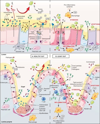
Table 1 Deleterious actions of pathogens in the gut and beneficial effects of S. boulardii CNCM I-745
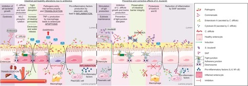
Figure 2 Proposed model for the effects of S. boulardii on intestinal permeability due to antibiotics.
Notes: Administration of antibiotics leads to multiple intestinal alterations. For example, antibiotic administration reduces all bacterial growth and this reduction leads to C. difficile growth and to the production of these toxins, which alter the formation of tight junctions. Intestinal permeability and the entry of pathogens into the lamina propria lead to different responses from the intestinal tissue: passage of electrolytes and water to the lumen, TNF-α production by macrophages leading to enterocyte apoptosis, and secretion of pro-inflammatory cytokines leading to inflammation. S. boulardii has a variety of preventive and corrective effects on antibiotic alterations: it stimulates the secretion of IgA directed against toxin A, and inhibits pathogen growth and toxin production. Besides, S. boulardii inhibits MLC phosphorylation and preserves the tight junction at the cell membrane. The restoration of intestinal barrier decreases the passage of electrolytes and water to the lumen. S. boulardii induces proteolysis of toxins A and B by secretion of serine protease (54 kDa) and secretes a factor named SAIF which exerts anti-inflammatory effects.
Abbreviations: C. difficile, Clostridium difficile; MLC, myosin light chain; NF-κB, nuclear factor-κB; S. boulardii, Saccharomyces boulardii; SAIF, S. boulardii anti-inflammatory factor.
Abbreviations: C. difficile, Clostridium difficile; MLC, myosin light chain; NF-κB, nuclear factor-κB; S. boulardii, Saccharomyces boulardii; SAIF, S. boulardii anti-inflammatory factor.