
Figures & data
Figure 1 Abdominal computed tomography scans showing the classical signs of mesentric panniculitis.
Notes: (A) Sign 1: axial view showing a well-defined mesenteric fatty mass (arrow) with mass effect on adjacent bowel loops (arrowheads). (B) Sign 2: axial view showing higher density of the mass (small circle) than the adjacent abdominal fat (large circle). (C) Sign 3: sagittal view showing the presence of blood vessels (arrowhead) and small lymph nodes (arrow) inside the mass. (D) Sign 4: coronal view showing the “halo sign” around the lymph nodes and mesenteric vessels (arrow). (E) Sign 5: axial view showing the presence of a hyperattenuating stripe around the mass, known as a pseudocapsule (arrow).
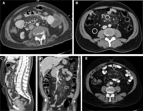
Table 1 Distribution of MP in both groups according to age and gender
Figure 2 Percentage of mesenteric panniculitis cases in both groups according to age group.
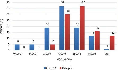
Table 2 Associated malignancies of the patients in Group 2
Table 3 CT characteristics of mesenteric panniculitis in both groups
Table 4 Total number of PC
Table 5 Measurement of the dimensions, volume, mass effect, fat density, and lymph nodes of the MP masses in both groups
Table 6 Risk factors for associated malignancy in patients with MP: RR with 95% CIs and ORs