
Figures & data
Figure 1 Abdominal Doppler ultrasonography with ascites, dilatation of portal vein, and low portal vein peak velocity.
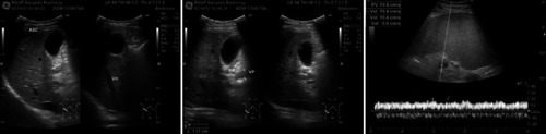
Figure 2 Contrast-enhanced abdominal CT scan with dilatation of portal vein with thrombus and dilatation of splenic vein.
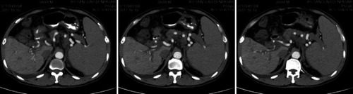
Figure 3 Esophagogastroduodenoscopy, esophageal varices grade 3 with the positive red color sign (left), esophagogastric junction (middle), and cardia stomach varices (right).
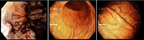
Figure 4 Step by step the operation. Left subcostal incision (A); splenectomy (B); transection and end-to-end anastomosis of the lower esophagus (C); devascularization of the upper 2/3 of the major gastric curvature (D); pyloroplasty (E); proximal splenorenal shunt (F).
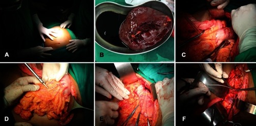
Figure 5 Esophagogastroduodenoscopy 2 months after surgery, esophageal varices grade 2 with the negative red color sign (left), cardia stomach edema (middle), and gastric snakeskin (right).
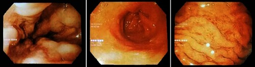
Table 2 Type of shunt techniques.Citation6,Citation12