
Figures & data
Figure 1 A high transsphincteric fistula.
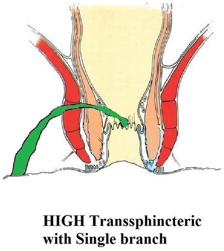
Figure 2 A high transsphincteric fistula with supralevator extension.
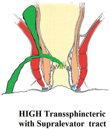
Figure 3 A suprasphincteric fistula.
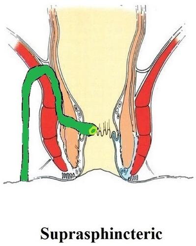
Figure 4 An extrasphincteric fistula.
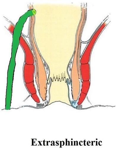
Figure 5 A 36-year-old male patient underwent surgery for a high intersphincteric fistula. Yellow arrows show the fistula tract/abscess. Left panel: preoperative MRI scans showing a high intersphincteric fistula. Middle panel: after 3 months of the first surgery, the fistula looked clinically healed with closed external opening (upper). However, the MRI scan revealed a large intersphincteric abscess (lower). The patient was operated on again. Right panel: MRI scan after 4 months of the second surgery shows complete radiological healing. The patient is doing well 18 months after the second surgery.
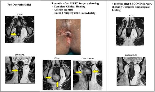
Table 2 Anal Fistula Classification
Table 1 Overview of Challenges in Managing Complex anal Fistulae and their Solutions