Figures & data
Figure 1 Acute cholecystitis (AC) with distended gallbladder, stones, mucosal hyper-enhancement and pericholecystic fluid noted on computed tomography (CT) scan.
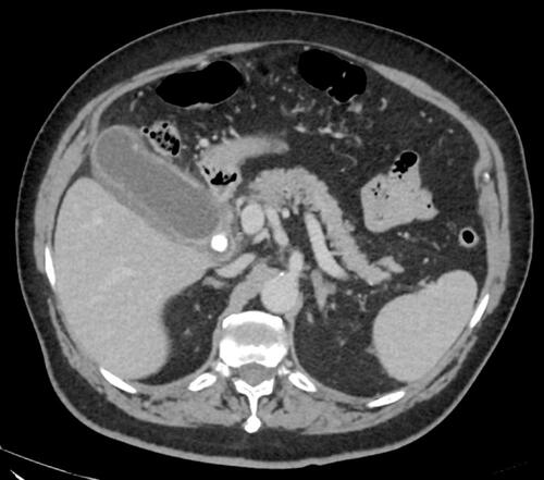
Figure 2 Graphic representation of the main interventional technique applied to perform gallbladder drainage in patients with acute cholecystitis (AC); (A) percutaneous transhepatic gallbladder drainage (PTGBD); (B) endoscopic transpapillary gallbladder drainage (ETGBD); (C) endoscopic ultrasound-guided gallbladder drainage (EUS-GBD) with electro cautery lumen-apposing metal stent (EC-LAMS).
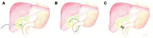
Table 1 Available Lumen Apposing Metal and Biflanged Stents
Figure 3 Endoscopic ultrasound-guided gallbladder drainage (EUS-GBD) with first flange opening of the electrocautery lumen-apposing metal stent (EC-LAMS).
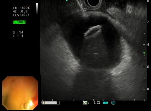
Figure 4 Endoscopic image obtained after electrocautery lumen-apposing metal stent (EC-LAMS) deployment in the gastric lumen.
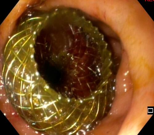
Figure 5 Fluoroscopic view of newly positioned electrocautery lumen-apposing metal stent (EC-LAMS) into the gallbladder lumen.
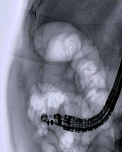
Figure 6 Computed tomography (CT) scan after 2 months of follow-up showing a cholecystoduodenostomy using electrocautery lumen-apposing metal stent (EC-LAMS).

Table 2 Comparison Between EUS-GBD and PTGBD
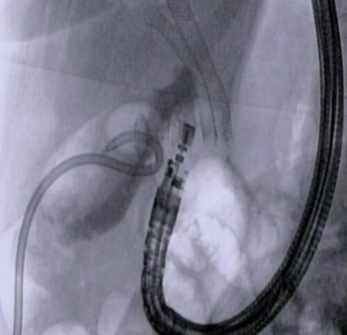
Figure 7 Fluoroscopic view of a conversion procedure, from percutaneous transhepatic gallbladder drainage (PTGBD) to endoscopic ultrasound-guided gallbladder drainage (EUS-GBD) with the positioned electrocautery lumen apposing metal stent (EC-LAMS) into the gallbladder lumen.