
Figures & data

Figure 1 Preferred reporting items for systematic reviews and meta-analyses (PRISMA) 2009 flow diagram: gallbladder hydatid cyst: literature search.
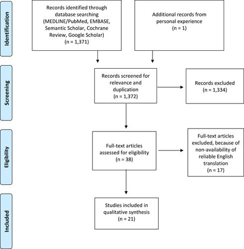
Figure 2 Computed tomography (CT) scan showing well-defined cystic lesion with internal septations and septal and peripheral calcification in segments IVa/IVb of the liver.
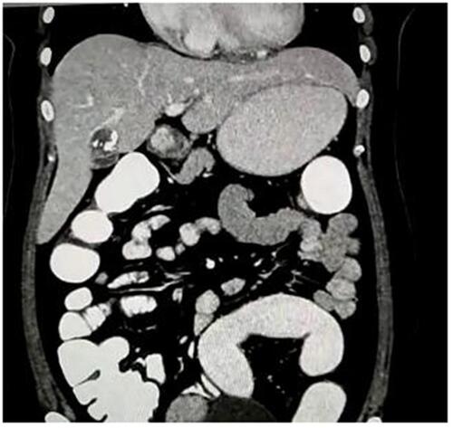
Figure 3 Inflamed deformed gallbladder after resection.
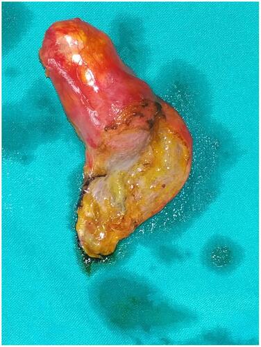
Figure 4 Opened gallbladder specimen with daughter hydatid cyst.
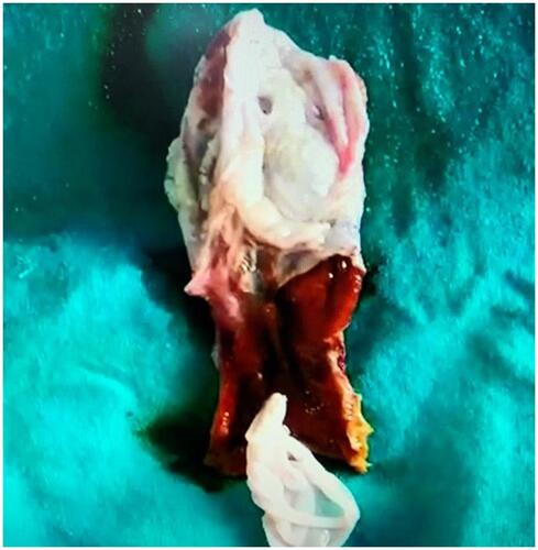
Table 1 Primary Gallbladder Hydatid Cyst: Clinical Features and Investigations
Table 2 Primary Gallbladder Hydatid Cyst: Management and Follow-Up
Table 3 Secondary Gallbladder Hydatid Cyst: Clinical Features and Investigations
Table 4 Secondary Gallbladder Hydatid Cyst: Management and Follow-Up