Figures & data
Figure 1 Experimental model.
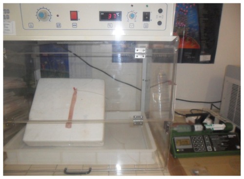
Figure 2 Perfusion sequences used for the evaluation of mucosal permeability and Esoxx effect.
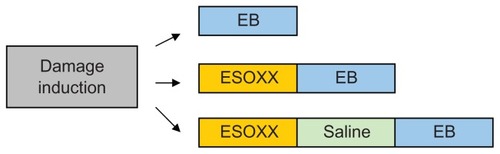
Table 1 Severity of histological damage induced with acidic and pepsin solutions
Figure 3 Representative examples of cross-sections of esophageal mucosa (H&E) treated with saline (A) or different damaging solutions (B–D). (A) No damage (grade 0). (B) Acid solution (60 minutes): mild damage (grade 1) extended throughout one or two epithelial layers. (C) Pepsin solution (30 minutes); moderate damage (grade 2) mainly localized on superficial layers. A disorganization of epithelial layers was observed along the tissue, with some intact areas and areas in which erosion interested from 30% to 50% of mucosal thickness. (D) Acid solution (90 minutes); severe damage (grade 3) and complete erosion of keratinic epithelial layers, with injury extending through more than 50% of epithelial stratified layer.
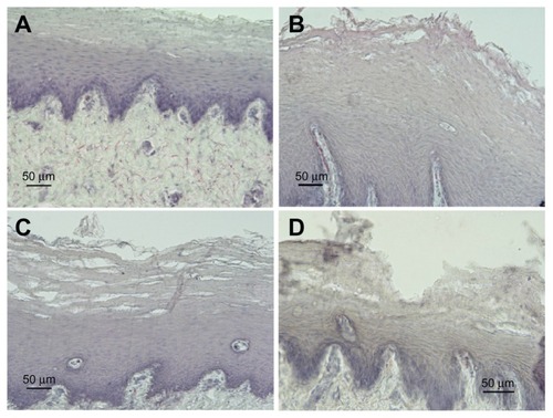
Figure 4 Representative examples of cross-sections of esophageal mucosa after EB perfusion. (A) No stain; undamaged mucosa + EB. (B) Weak stain; damaged mucosa (acid solution 90 minutes) + EB. (C) Strong stain; damaged mucosa (pepsin solution 60 minutes) + EB. (D) No stain; damaged mucosa (acid solution 90 minutes) + Esoxx + EB.
Abbreviation: EB, Evans blue dye.
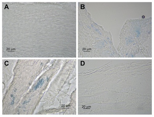
Table 2 Evans blue (EB) staining on control tissue and damaged mucosa with or without Esoxx application