Figures & data
Figure 1 Abdominal and pelvic CT scan of a 75-year-old man showing a perforation of the anterior wall of the rectum.
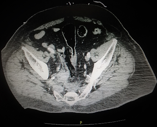
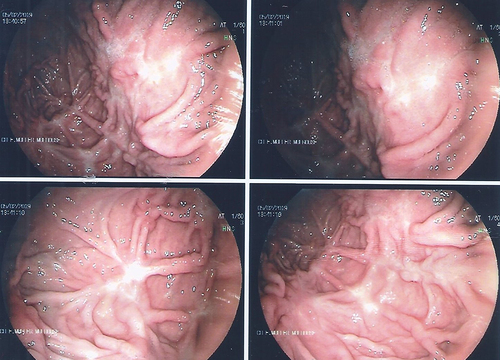
Figure 2 Upper gastrointestinal endoscopy showed suspicious ulcerated lesions along the large anterior gastric tuberosity, surrounded by a budding mucosa.
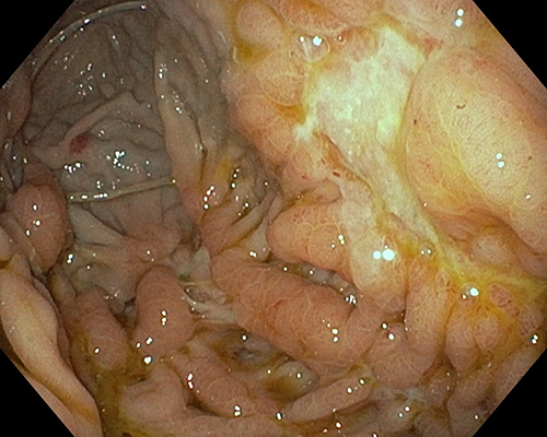
Figure 3 Recto-sigmoidoscopy of a 75-year-old man showing uncommon fistulous orifice with leakage on the anterior rectal wall.
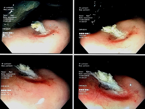
Figure 4 Histopathology of the gastric biopsies had shown a typical of Muchorales image, suggestive mucormycosis.
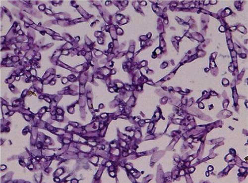
Figure 5 Upper gastrointestinal endoscopy 4 weeks after liposomal amphotericin B, showing numerous scarred stellar lesions sometimes with retraction of the greater gastric tuberosity and anterior wall.