Figures & data
Table 1 Clinical outcomes of POEM for esophageal achalasia
Table 2 Clinical outcomes of endoscopic treatment using submucosal tunneling technique for GI subepithelial tumors
Table 3 Clinical outcomes of tissue sampling methods for GI subepithelial tumors
Figure 1 Endoscopic findings using submucosal tunneling technique of tumor enucleation in a SET located in the esophagogastric junction.
Notes: (A) Endoscopic finding showed a flat elevated subepithelial lesion in the esophagogastric junction, which might lead to stenosis by enlargement of the tumor in the future. (B) After making an entry site at 2 cm from the tumor’s edge, a submucosal tunnel was created by submucosal dissection using a needle-knife form. After the tumor was identified and exposed in the tunnel, submucosa around the tumor was dissected. A white-colored tumor in the submucosal layer was enucleated completely. Finally, the entry site was sutured completely with hemoclips. (C) Macroscopic image of the resected specimen (20×12 mm). IH findings resulted in a gastric leiomyoma. (D) Follow-up endoscopy 2 weeks after operation revealed no tumor or esophagogastric stenosis.
Abbreviations: SET, subepithelial tumor; IH, immunohistological.
Abbreviations: SET, subepithelial tumor; IH, immunohistological.
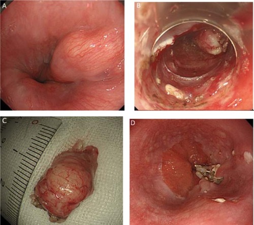
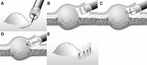
Figure 2 TBB procedure using submucosal endoscopy with the mucosal flap method for GI subepithelial tumors.
Notes: (A) Endoscopic submucosal dissection: a 10 mm opening flap was created by mucosal incision and submucosal dissection. (B) SEMF: a short tunnel approaching the tumor was created by additional submucosal dissection. (C) Bloc biopsy: a bloc specimen, measuring 5×5×2 mm, was obtained using a needle-knife and the cutting mode of the electrosurgical unit. (D) Tissue collection into a transparent cap. Specimen was removed into a long attachment, using grasping forceps. (E) Clip closure of flap. Opening flap was completely closed with hemoclips.
Abbreviations: TBB, tunneling bloc biopsy; GI, gastrointestinal; SEMF, submucosal endoscopy with mucosal flap.
Abbreviations: TBB, tunneling bloc biopsy; GI, gastrointestinal; SEMF, submucosal endoscopy with mucosal flap.