
Figures & data
Figure 1 Dysregulation of coagulation, complement, and contact cascades in hereditary angioedema.
Abbreviations: HAE, hereditary angioedema; MASP-2, mannose-binding lectin-associated serine protease 2.
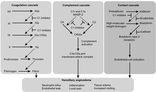
Figure 2 Sites affected by angioedema in patients with clinical symptoms of hereditary angioedema.
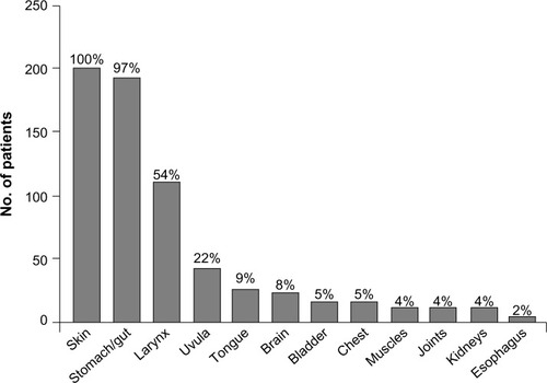
Figure 3 Phases and time course for typical abdominal pain attacks in patients with hereditary angioedema.
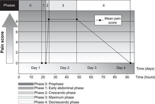
Table 1 Differential diagnosis of intestinal angioedema
Figure 4 Abdominal computed tomography scan of patient with hereditary angioedema showing thickening of the small bowel (stacked-coin appearance) due to angioedema.
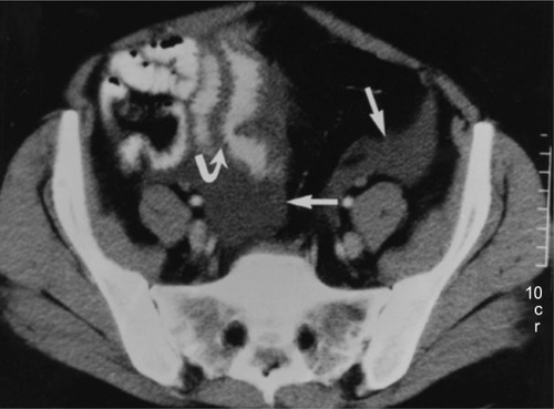
Figure 5 Diagnostic algorithm for hereditary angioedema.
Abbreviations: AE, adverse event; ACE, angiotensin-converting enzyme; C1-INH, C1 esterase inhibitor; C1q, complement component 1, q subcomponent; HAE, hereditary angioedema.
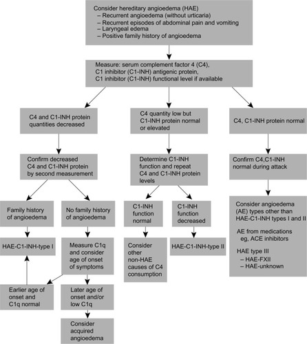
Table 2 Hereditary Angioedema International Working Group consensus guidelines for the management of hereditary angioedema
Table 3 Specific agents for hereditary angioedema (HAE)