
Figures & data
Table 1 Characteristics and presentations of patients
Figure 1 Examples of foreign bodies.
Notes: Coins (A) were the most common foreign bodies. Pins (B) were common in females and commonly seen piercing the antrum. A surgical towel after cholecystectomy (C) was encountered in only one patient. A fleshy meat bolus (D) was commonly trapped at esophageal stricture.
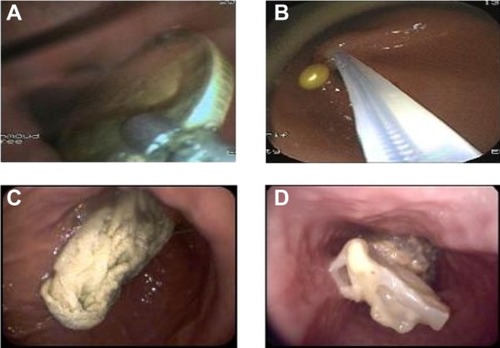
Figure 2 Surgical removal (A) of a stomach shaped (B) bezoar in a mentally disabled female.
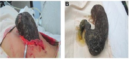
Table 2 Types of trapped foreign bodies
Figure 3 X-ray was used to localize the foreign body (coin; arrow) in some cases (A), and computed tomography (B) was used to describe the huge gastric bezoar (arrow) in the case of a mentally disabled patient.
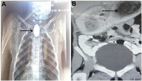
Table 3 Site of trapped foreign bodies
Table 4 Treatment outcomes