Figures & data
Figure 1 Gastric antral vascular ectasia on initial upper endoscopy.
Notes: (A, B) Different views demonstrating the same findings.
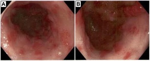
Figure 2 Immediately post radiofrequency ablation.
Notes: (A, B) Different views demonstrating the same findings.
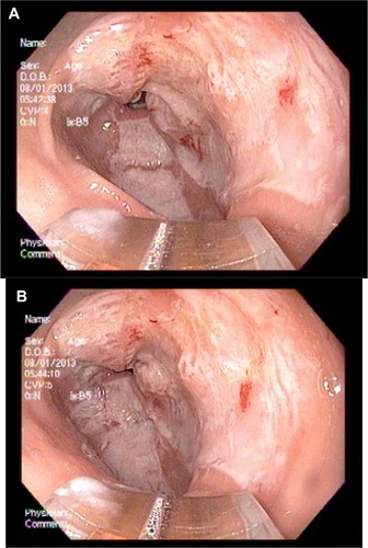
Figure 3 Upper endoscopy 1 year after radiofrequency ablation.
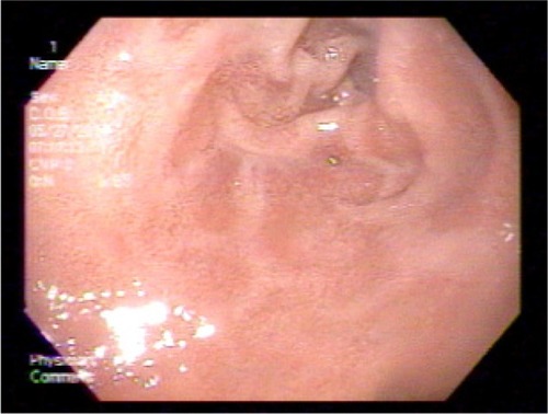
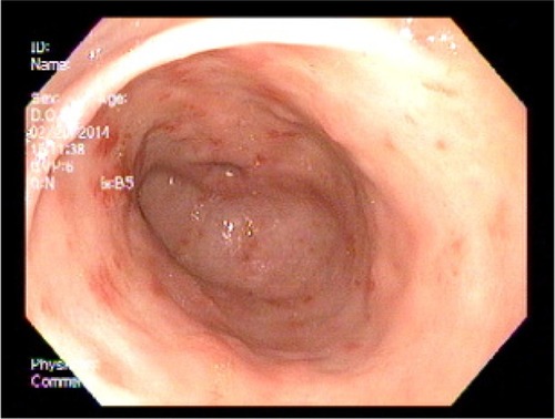
Figure 4 Upper endoscopy showing the typical longitudinal red stripes in the antrum radiating out from the pylorus, consistent with gastric antral vascular ectasia.
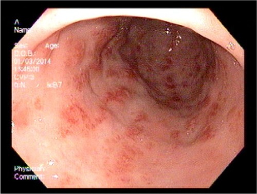
Figure 5 Longitudinal, erythematous stripes can again be seen radiating in a spoke-like fashion from the pylorus to the antrum.
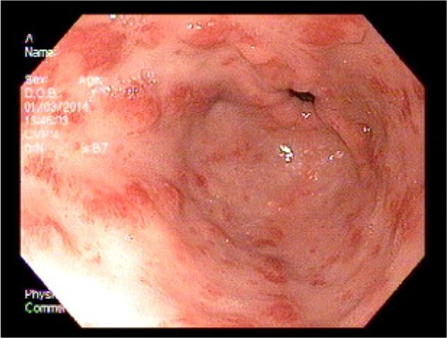
Figure 6 Upper endoscopy immediately after cauterization with the gold probe hemostasis catheter.
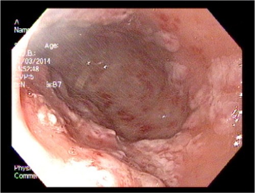
Figure 7 Upper endoscopy before therapeutic intervention with radiofrequency ablation.