
Figures & data
Figure 1 The 3D-Transit system
Notes: (A) Capsule containing an electromagnet and a battery (inset). The abdominal belt contains the 3D-Transit receiver and is worn by the subjects throughout the examination. Thoracic belt (blue) and an accelerometer inside the detector record artifacts owing to breathing and posture changes. The ruler is measuring in centimeters. (B) 3D-Transit visualization and analysis software. Changes in position and orientation of the capsules with respect to the receiver reflect gastrointestinal contractile activity and progression. Coordinates x, y, and z display capsule movements in a frontal, sagittal, and transversal plane, respectively. The Φ,θ coordinates describe capsule rotation, reflecting the contraction frequency (arrows mark individual contractions). The inset shows the actual movement of the capsule, which corresponds to the changes in the coordinates. The x-axis represents time.
Abbreviations: R, respiration; sec, seconds.
Abbreviations: R, respiration; sec, seconds.
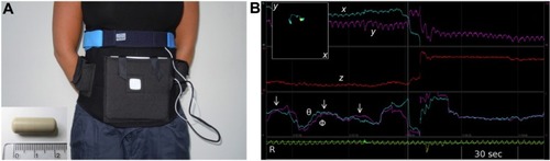
Figure 2 Data synchronization.
Notes: Posture changes obtained through the accelerometer inside the 3D-Transit receiver (red, green, and blue lines) are associated with an increased heart rate achieved through the portable sleep monitor. This way data were easily time-linked (ovals).
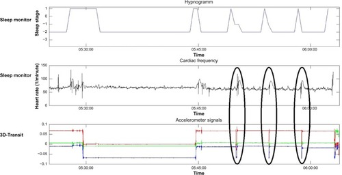
Table 1 Sleep distribution and efficiency
Table 2 Gastric contractions and basal colonic activity during sleep and nocturnal awakenings