Figures & data
Table 1 Summary of the GLUT family proteins and their characteristics
Figure 1 Amino acid sequences alignments of the GLUT family of proteins.
Notes: Transmembrane domains are highlighted in yellow based on the new GLUT1 crystal structure. Highly conserved residues are colored in red. Residues highlighted in blue in TM10 are believed to be the cytochalasin B recognition/binding sites. Residues highlighted in blue in TM7 are believed to be the critical hydrophobic residues responsible for substrate selectivities.
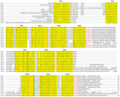
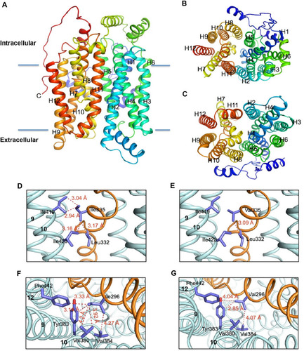
Figure 2 Molecular models of the human hGLUT9 and hGLUT5 transporters comparing possible hydrophobic interactions.
Notes: (A) Computer model of hGLUT9a based on the human hGLUT1 crystal structure. (B and C) Views from the intracellular face and extracellular face. (D) Potential interactions of I335 indicate a hydrophobic network with residues within TM10. (E) Structural model of the mutant SLC2A9 I335V was generated, demonstrating that the intricate linkage to helix 10 is disrupted when I335 is converted to Val. (F) In SLC2A5, Ile296, the equivalent of Ile335 in SLC2A9, forms a more extensive hydrophobic cluster with neighboring residues in TM10 and TM12. (G) Structural model of the mutant SLC2A5 I296V, highlighting loss of the hydrophobic network, which subsequently leads to alteration in substrate specificity. Copyright © 2015 American Society for Biochemistry and Molecular Biology. Adapted from Long W, Panwar P, Witkowska K, et al. Critical roles of two hydrophobic residues within human glucose transporter 9 (hSLC2A9) in substrate selectivity and urate transport. J Biol Chem. 2015;290: 15292–15303.Citation61