Figures & data
Table 1 The characteristics of the three cases, before and after treatment
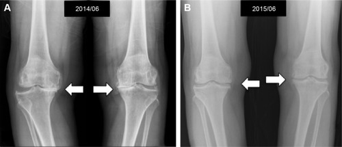
Figure 1 Standard weight-bearing knee X-rays of a 77-year-old female before (A) and 8 months after (B) one course of intra-articular PRP in association with HA injection, showing the increase in medial joint space (white arrows).
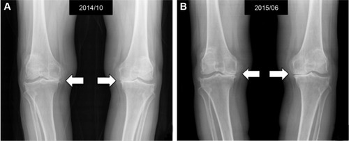
Figure 2 Standard weight-bearing knee X-rays of a 69-year-old female before (A) and 15 months after (B) one course of intra-articular PRP in association with HA injection, showing significant improvement in the narrowing joint space (white arrows).
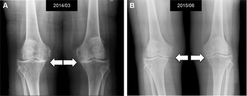
Figure 3 Standard weight-bearing knee X-rays of a 76-year-old female before (A) and 1 year after (B) two courses of intra-articular PRP in association with HA injection, showing more significant bone spur growth and the cartilage remains at a healthy size with the normal joint space (white arrows).