Figures & data
Table 1 Characteristics of study participants
Table 2 Application of 18F-FDG PET/CT and CA19-9 in differentially diagnosing PC from CMFP
Figure 1 Images of a male participant, 69 years.
Notes: Computed tomography showed that there was a suspicious lesion in his pancreas. His carbohydrate antigen 19-9 level was 51.68. 18F-FDG PET/CT showed that there was no lesion with increased 18F-FDG uptake in his pancreas. Finally, he was diagnosed with chronic mass-forming pancreatitis. (A) Pancreatic head lesion in computed tomography; (B) pancreatic head lesion in 18F-FDG PET/CT; (C) pancreatic duct dilatation in computed tomography; and (D) pancreatic duct dilatation in 18F-FDG PET/CT.
Abbreviations: PET, positron emission tomography; CT, computed tomography.
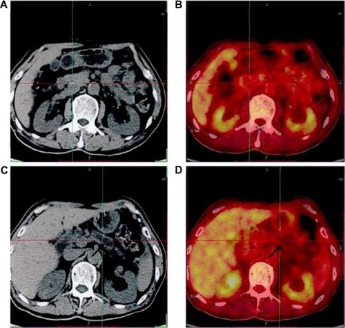
Figure 2 Images of a female participant, 66 years.
Abbreviations: PET, positron emission tomography; CT, computed tomography.
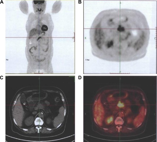
Figure 3 Scatter plot for participants with chronic mass-forming pancreatitis between standardized uptake value maximum and carbohydrate antigen 19-9 levels.
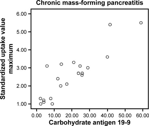
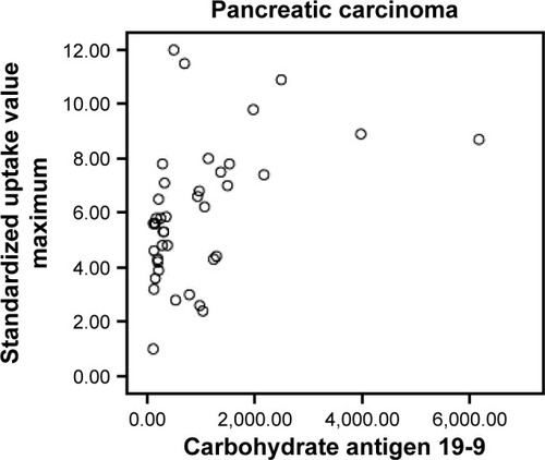
Figure 4 Scatter plot for participants with pancreatic carcinoma between standardized uptake value maximum and carbohydrate antigen 19-9 levels.