
Figures & data
Table 1 Participant characteristics in the two groups
Figure 1 A male patient (63 years old) with type III pilon fracture.
Notes: (A) Preoperative anteroposterior X-ray; (B) Preoperative anteroposterior and lateral X-ray; (C) Postoperative anteroposterior X-ray; (D) Postoperative anteroposterior and lateral X-ray.
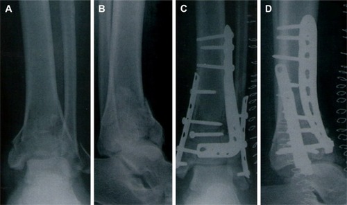
Table 2 Clinical value of the 2 surgeries