
Figures & data
Figure 1 Sonography, injection, and blinding for injector.
Note: The syringe was covered by a sterile opaque white paper (left picture); flexor retinaculum (FR), median nerve (MN), and the injectate (INJ) for dissection are shown in the middle picture; covering the needle at the time of withdrawal (right picture).
Abbreviations: FT, flexor tendon; CB, carpal bone.
Abbreviations: FT, flexor tendon; CB, carpal bone.

Figure 2 Study flowchart.
Note: Group I received 80 mg triamcinolone (2 mL) and 1 mL of 2% lidocaine; Group II received 40 mg triamcinolone (1 mL), 1 mL of 2% lidocaine, and 1 mL normal saline; Group III received 1 mL of 2% lidocaine and 2 mL normal saline.
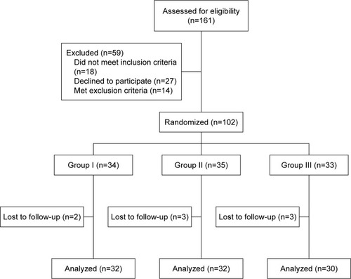
Table 1 Patients’ demographics
Table 2 Outcome measures at baseline and follow-up
Table 3 Between-group analyses