Figures & data
Figure 1 Bran magnetic resonance imaging (MRI) showed some abnormal T2 hyperintense signals involving in the splenium and the body of corpus callosum, midbrain and bilateral internal capsule, with slight contrast enhancement (C).
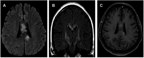
Figure 2 Cervical MRI indicated T2 hyperintense lesions involving C3-T5.
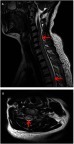
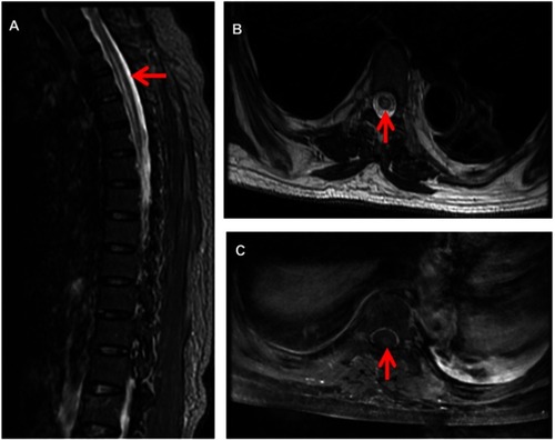
Figure 3 Contrast enhanced thoracic MRI showed T2 hyperintense lesions extending from T7-T12, with slight contrast enhancement (C).