
Figures & data
Figure 1 Placement of electrodes used for NMES application.
Abbreviation: NMES, neuromuscular electrical stimulation.
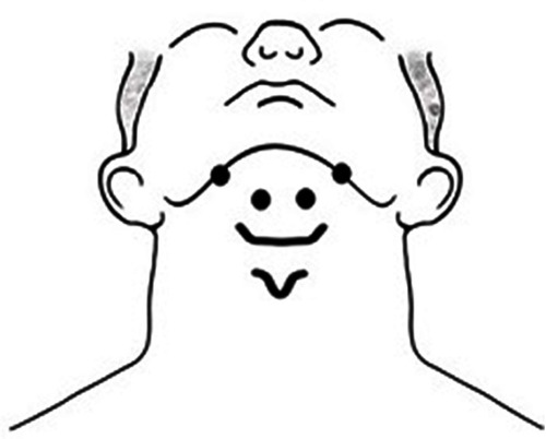
Figure 2 Placement of sEMG electrodes on the supra- and infrahyoid muscles.
Abbreviation: sEMG, surface electromyography.
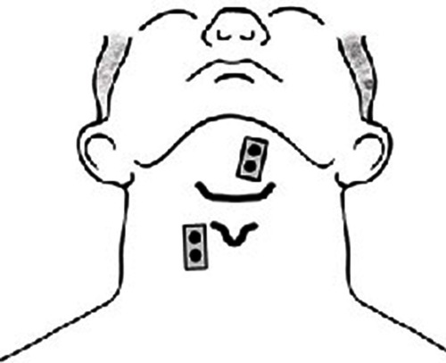
Figure 3 (A) Raw EMG signal of the suprahyoid muscles (above trace) and the infrahyoid muscles (below trace) during swallowing. (B) The rectified EMG signals of the suprahyoid muscles (above trace) and the infrahyoid muscles (below trace). Abbreviation: EMG, electromyographic.
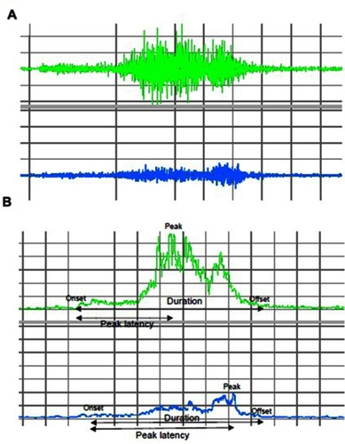
Figure 4 Subject dropout.
Abbreviations: HLE, head lift exercise; NMES, neuromuscular electrical stimulation.
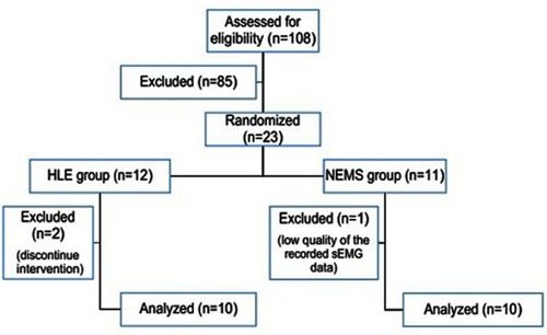
Table 1 The mean ± standard deviation of sEMG parameters of the suprahyoid muscle activity before and after treatment
Table 2 The Mean±standard deviation of sEMG parameters of the infrahyoid muscle activity before and after treatment