
Figures & data
Figure 1 (A) Brain diffusion-weighted-MRI of the proband patient. (B) FDG PET imaging of proband patient.
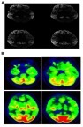
Figure 2 Family tree of the patient.
Abbreviation: CJD, Creutzfeldt–Jakob disease.
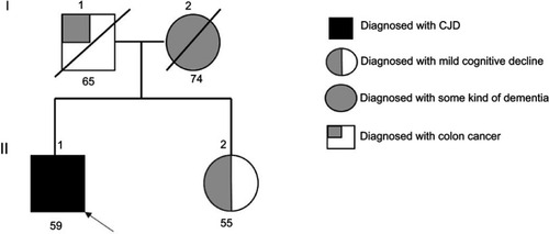
Figure 3 Next generation sequencing data for a patient with PRNP Tyr225Cys, verified by standard sequencing.
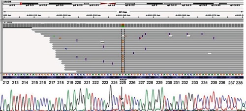
Figure 4 (A) ExPASY analysis of PRNP Tyr225Cys compared with normal PrP protein. (B) Comparison of normal PrP proteins with Tyr225 and mutant Cys225 in terms of distance from Met166. (C) In silico prediction of PRNP Tyr225 and Cys225. Helix-3 in prion proteins may be more flexible in the case of Cys225 due to the smaller size of cysteine.
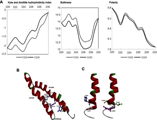
Figure S1 EEG data of the patient.
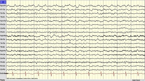
Table 1 Examples of mutations, observed in atypical CJD cases
Table 2 Mutations, located in the C-terminal region of 3rd helix of prion protein
Table S1 SF tapping profile of the proband patient