
Figures & data
Table 1 Patient Demographics and Operative Characteristics in PELD Group and Fenestration Group
Table 2 General Clinical Results in the PELD Group and Fenestration Group
Figure 1 Clinical outcomes before and after endoscopic decompression at different follow-up time points in PELD group and fenestration group. (A) Visual analog scale (VAS) scores for back pain. (B) VAS scores for leg pain. (C) Japanese Orthopaedic Association Score (JOA) scores.
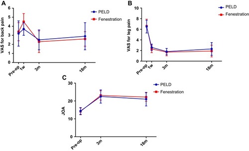
Figure 2 Outcomes of 36-Item Short-Form Health Survey (SF-36) before and at the final follow-up.
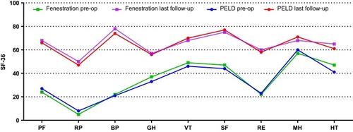
Figure 3 Satisfaction rates according to the modified Macnab criteria in PELD group (A) and fenestration group (B) at the final review (18 months) post-surgery.
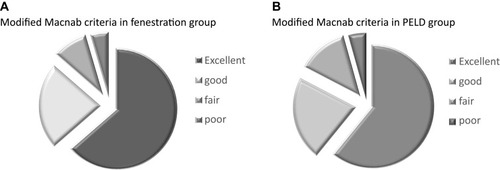
Figure 4 An 81-year-old female patient with lumbar lateral recess stenosis (LRS) who received PELD. (A) Preoperative magnetic resonance images (MRI) showing severe lateral recess stenosis with LRS at the left L4-5 level (yellow arrowhead). (B) Postoperative MRI showing a thorough nerve decompression (yellow arrowhead).
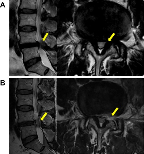
Figure 5 A 76-year-old male patient with lumbar lateral recess stenosis (LRS) who received fenestration. (A) Preoperative computed tomography (CT) images showing severe lateral recess stenosis with LRS at the left L4-5 level (yellow arrowhead). (B) Postoperative CT images showing a thorough nerve decompression (yellow arrowhead).
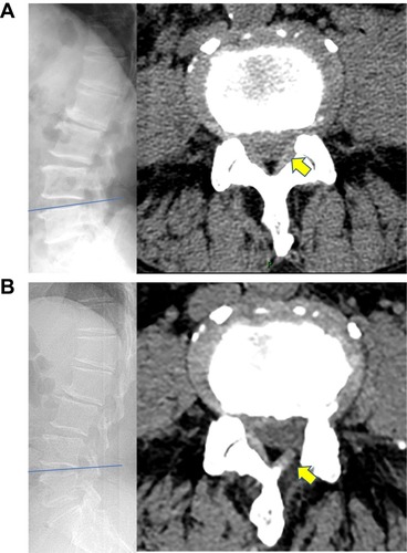
Table 3 Comparison of Clinical Results in PELD Group and Fenestration Group with Other Conventional Open Decompression Surgery for Lumbar Stenosis