
Figures & data
Figure 1 (A and B) Enhanced angiographic CT examination in and (C and D) maximum intensity projection technique showed a complex tangle of dilated and tortuous vessels of the right-sided temporoparietooccipital regions and posterior condylar area (arrowheads).
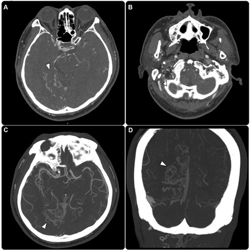
Figure 2 (A) Axial T2 weighted image demonstrates fine tortious dural vessels at the right parietooccipital and temporal region (arrowheads). (B and C) Susceptibly weighted image (SWI) showed engorged dural vessels with multifocal intraparenchymal hemorrhage in the right temporal and occipital area (arrows). (D) Arterial spin labeling (ASL) showed an increased blood pool in the right occipital and temporal region, resembling venous congestion.
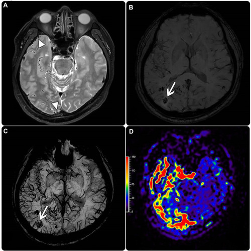
Figure 3 (A) Lateral cerebral angiogram selectively through the left external carotid artery (arrowhead) showed Torcula dAVF (asterisk) and posterior condylar vein (arrow). (B and C) Cerebral angiogram lateral view and 3D reconstruction of the right external carotid artery injection respectively showed posterior condylar vein (arrow), lateral sinus dAVF (asterisk) and the left external carotid artery (arrowhead). (D) AP view cerebral angiogram through the left external carotid artery showed Torcula dAVF (arrowhead). (E) Three dimensional both ECA angiogram (fused image) visualizes Torcula dAVF, and lateral sinus dAVF (arrowheads).
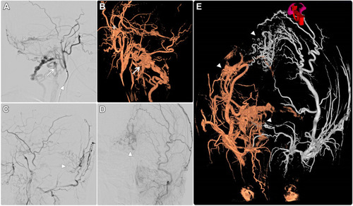
Figure 4 (A) The selective roadmap of the lower skull part shows super-selection of paravertebral venous plexus reaching to posterior condylar vein dAVF (arrow). (B) Platinum coils were deployed at the posterior condylar vein dAVF (arrows). (C) The final run shows the total obscuring of posterior condylar vein AVF (asterisk). (D and E) Post-operative computed tomography images reveal complete coil material at the location of posterior Condylar vein dAVF, with embolic material obscuring the lateral sinus Davf (arrowheads).
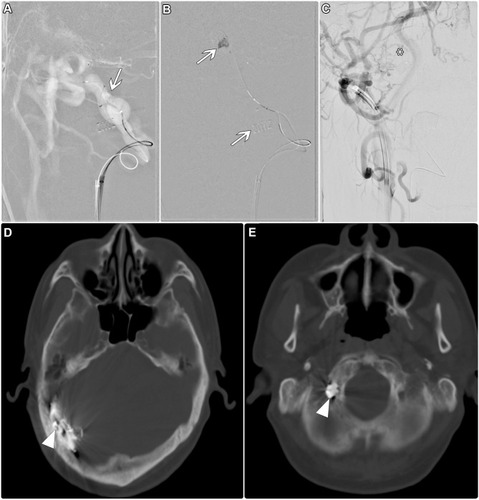
Table 1 A Summary of Reported Cases of dAVFs Involving the Posterior Condylar Vein