Figures & data
Figure 1 Placement of the electrodes for surface electromyography measurement.
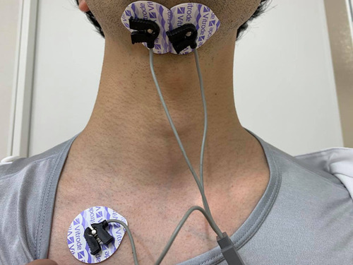
Table 1 Characteristics of Study Participants
Table 2 Comparison of sEMG Parameters and Lingual Pressure in the Non-Dysphagic and Sarcopenic Dysphagic Groups
Table 3 Diagnostic Accuracy of sEMG Parameters and Cut-Off Values for Sarcopenic Dysphagia
Figure 2 Submental surface electromyography (sEMG) patterns in patients without and with dysphagia. (A) sEMG pattern in a patient without dysphagia. (B) sEMG pattern in a patient with sarcopenic dysphagia. Left-most blue vertical line, swallow onset; middle blue vertical line, maximum amplitude point; right-most blue vertical line, end of the swallowing activity.
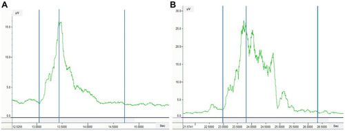
Table 4 Association Between Duration, Amplitude, and Sarcopenia-Related Factors
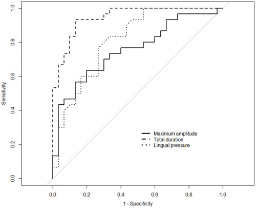
Figure 3 Receiver operating characteristic curve for the diagnosis of sarcopenic dysphagia.