Figures & data
Figure 1 CT images showed “bamboo-like changes” in the spine, and T12 showed a fresh three-column fracture (red arrow) (A) A pseudarthrosis with marked sclerosis at T12, and the upper part of the T12 vertebral body was damaged irregularly (red arrow) (B) The lesion involved the intervertebral space and the lower part of the T11 vertebral body, with pseudarthrosis and obvious hyperplasia (red arrow) (C) The scope of the lesions had extended, with severe thoracolumbar kyphosis and spinal canal stenosis (red arrow) (D) The position of the pedicle screw was well fixed, the kyphosis was obviously corrected, and the intervertebral bone graft was adequate (red arrow) (E) AL segmental bone grafts have good fusion (red arrow) (F).
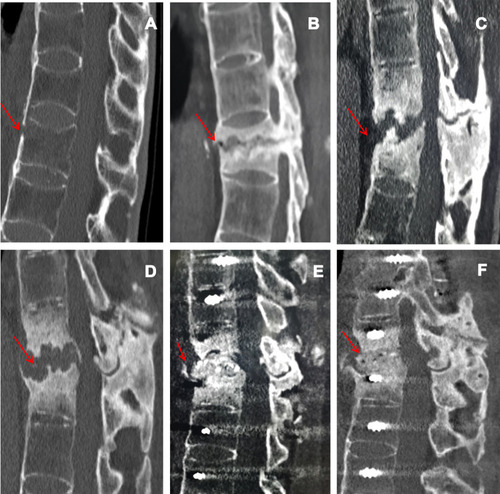
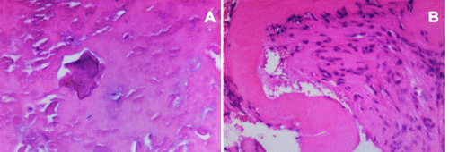
Figure 2 Histopathologic analysis showed fibrocartilage tissue calcification and degeneration, necrotic intervertebral disc tissue, fibrovascular hyperplasia, and focal aggregation of inflammatory cells (A and B).