
Figures & data
Table 1 Type and Frequency of Comorbidities
Table 2 Preoperative Functional Status According to the KPS Scale
Figure 1 Preoperative contrast-enhanced T1-weighted MR images in the axial (A) and coronal (B) planes of a 71-year-old woman operated on for a parasagittal meningioma. Last follow-up MRI examination obtained after Simpson grade III resection of a fibroblastic meningioma (WHO grade I) showed no recurrence in contrast-enhanced T1-weighted images in the axial (C) and sagittal (D) planes.
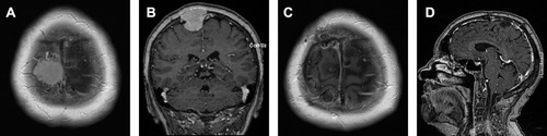
Figure 2 Preoperative contrast-enhanced T1-weighted MR image in the axial plane (A) of an 82-year-old woman operated on for a convexity meningioma. Last follow-up MRI examination obtained after Simpson grade I resection of an atypical meningioma (WHO grade II) with no postoperative radiotherapy showed no recurrence in the native T1-weighted image in the axial plane (B) and in contrast-enhanced T1-weighted images in the axial (C) and coronal (D) planes.
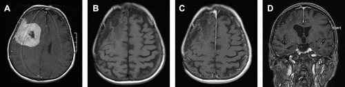
Figure 3 Preoperative MRI examination in contrast-enhanced T1-weighted axial (A) and T2-weighted coronal (B) planes of a convexity meningioma in a 72-year-old woman with Simpson grade I resection of an atypical meningioma (WHO grade II). No postoperative radiotherapy was used. MRI examination performed 18 months after surgery revealed a small recurrence shown in the axial contrast-enhanced T1-weighted image (C). Cyberknife radiotherapy was used. MRI examination performed 42 months after surgery showed further tumor growth (D) and Cyberknife radiotherapy was reapplied. The patient was alive at the last follow-up contact (50 months post-surgery).
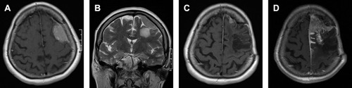
Table 3 Neurological Morbidity, Overall Morbidity and Mortality in Relation to the Simpson Grade of Removal
Table 4 The Relationship Between the Grading Scales’ Scores and the Incidence of Neurological Morbidity, Overall Morbidity, and Mortality
Table 5 The Course of Patients with Tumor Recurrence