Figures & data
Table 1 Compare Baseline Characteristics and Surgical Results of the Two Groups
Figure 1 3D-printed artificial vertebral body: oblique (A), lateral (B), and superior (C) views.
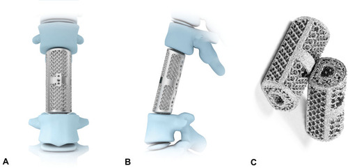
Figure 2 Measuring method of local kyphotic angle (LKA), anterior vertebral height (AVH), and posterior vertebral height (PVH).
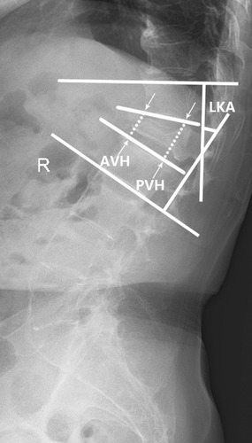
Table 2 Compare Preoperative and Final Follow-Up ASIA Between the Two Groups
Figure 3 Clinical and radiographic outcomes. (A–E) *P<0.05 compared with the preoperative data. (A–C) †P>0.05 compared with the 1-month postoperative data, ‡P<0.05 compared with the 1-month postoperative data, §P>0.05 compared with the 6-month postoperative data. (D) †P>0.05 compared with the 1-month postoperative data; ‡P>0.05 compared with the 6-month postoperative data. (E) †P<0.05 compared with the 1-month postoperative data.
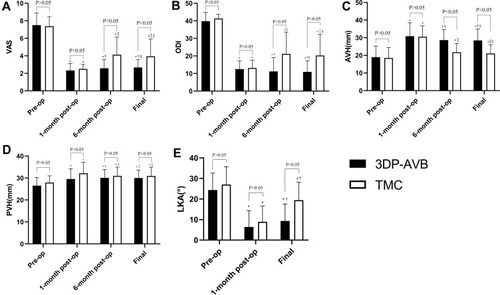
Table 3 Compare the Subsidence Outcomes and Subsidence Grade Between the Two Groups
Table 4 Radiographic and Clinical Outcomes According to Different Subsidence Grades
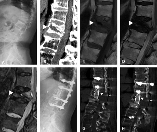
Figure 4 An 82-year-old female patient experienced Kümmell’s disease complicated by neurological deficits and used a 3D-printed artificial vertebral body (3DP-AVB). (A) T11 collapsed fracture was shown in preoperative X-rays. (B and C) Coronal and sagittal CT showed an intravertebral cleft (IVC) (white triangle). (D–F) MRI showed a decreased signal in IVC (white triangle). (G) Intraoperative images. (H and I) Immediate postoperative X-rays showed that a 3DP-AVB was implanted. (J) Postoperative sagittal T2-weighted MRI showed a sufficient spinal cord decompression. (K and L) Coronal and sagittal CT showed that no subsidence occurred at the final follow-up.
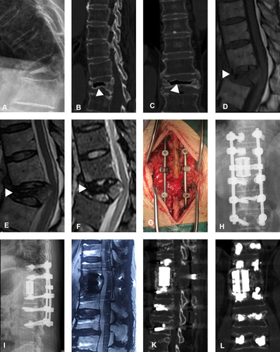
Figure 5 The 3D-printed artificial vertebral body (3DP-AVB) was used in a 75-year-old female patient with stage III Kümmell’s disease. (A) Preoperative X-rays showed T12 collapsed fracture and thoracolumbar kyphosis. (B and C) An intravertebral cleft sign (white triangle) was shown in preoperative CT scans. (D–G) Postoperative and final follow-up plain radiographs showed no cage subsidence and kyphosis recurrence.
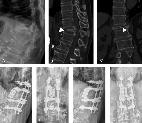
Figure 6 A titanium mesh cage (TMC) for stage III Kümmell’s disease. (A and B) Preoperative radiographs and CT scans revealed collapsed fracture and kyphosis at L1 with an intravertebral cleft (IVC) sign (white triangle). (C–E) Preoperative MRI showed a decreased signal of IVC (white triangle). (F) No subsidence in immediate postoperative plain radiographs. (G and H) Final follow-up CT scans showed kyphosis recurrence with TMC subsidence (white triangle).