Figures & data
Table 1 Primer Sequences Used for qPCR
Figure 1 The effect of PPF treatment on rats with MIRI and its effect on HOTAIR and miR-17-5p expression. (A) CK-MB in the serum of the rats in each group was detected with ELISA (N=3). (B) LDH in the serum of the rats in each group was detected with ELISA (N=3). (C) qRT-PCR was used to detect the expression level of HOTAIR in the heart tissue of the rats (N=3). (D) MiR-17-5p expression in heart tissue of the rats was detected by qRT-PCR. (E and F) Western blot was used to detect by the expression of p-STAT3 and STAT3 in heart tissue of the rats. ** P<0.01 and *** P<0.001.
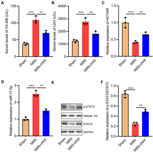
Figure 2 The effects of PPF on HOTAIR, miR-17-5p, and STAT3 expressions in H9c2 cells. (A and B) The expression levels of HOTAIR and miR-17-5p in H9c2 cells were detected by qRT-PCR (N=3). (C and D) Western blot was used to detect the expressions of STAT3 and p-STAT3 in H9c2 cells (N=3). * P<0.05, ** P<0.01 and *** P<0.001.
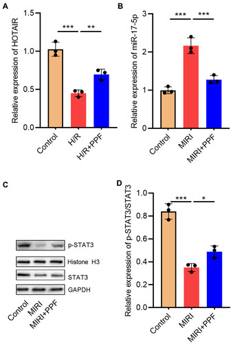
Figure 3 HOTAIR adsorbed miR-17-5p and repressed its expression in H9c2 cells. (A) StarBase database analysis revealed that there was a potential binding site between HOTAIR and miR-17-5p. (B) Dual-luciferase reporter analysis showed that miR-17-5p could inhibit the luciferase activity of HOTAIR-WT, but could not inhibit that of HOTAIR-MUT reporter (N=3). (C) RIP experiments indicated the direct interaction between HOTAIR and miR-17-5p (N=3). (D and E) qRT-PCR results showed that HOTAIR negatively regulated miR-17-5p expression in H9c2 cells (N=3). *** P<0.001.
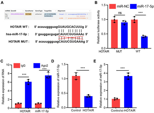
Figure 4 H/R treatment regulated miR-17-5p and STAT3 expressions via HOTAIR. (A) Empty plasmids and HOTAIR overexpression plasmids were transfected into H9c2 cells treated with H/R, respectively, and the expression of miR-17-5p was detected by qRT-PCR (N=3). (B and C) The expressions of STAT3 and p-STAT3 in H9c2 cells were detected by Western blot (N=3). (D) Control siRNA and HOTAIR siRNA were transfected into H9c2 cells treated with H/R and PPF, and the expression of miR-17-5p was detected by qRT-PCR (N=3). (E and F) The expressions of STAT3 and p-STAT3 in H9c2 cells were detected by Western blot (N=3). ** P<0.01 and *** P<0.001. ns, P>0.05.
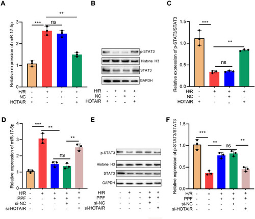
Figure 5 The effects of HOTAIR, STAT3, and PPF on H/R-induced apoptosis of H9c2 cells. (A–G) Apoptosis of H9c2 cells was detected by flow cytometry after HOTAIR or STAT3 was selectively regulated (N=3). ** P<0.01, and *** P<0.001. ns, P>0.05.
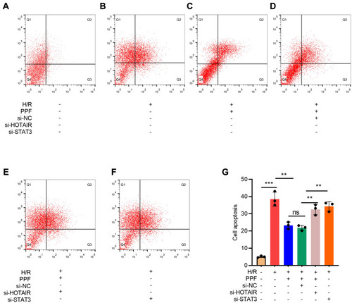
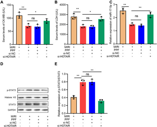
Figure 6 The effects of HOTAIR on the protective function of PPF in MIRI rats. (A) CK-MB in the serum of the rats in each group was detected with ELISA (N=3). (B) LDH in the serum of the rats in each group was detected with ELISA (N=3). (C) The expression level of miR-17-5p in the myocardial tissue of rats was detected by qRT-PCR (N=3). (D and E) Western blot was used to detect the expressions of STAT3 and p-STAT3 protein in cardiac tissue of the rats (N=3). * P<0.05, ** P<0.01. ns, P>0.05.