Figures & data
Table 1 Sequences of Primers Used in RT-qPCR
Figure 1 Patients with osteoporosis had increased microRNA-151a-3p level and reduced BMD. (A) MicroRNA-151a-3p level and BMD in patients with osteoporosis and control group were detected. (B) The correlation between microRNA-151a-3p and BMD was analyzed by Pearson’s correlation coefficient. **P < 0.01.
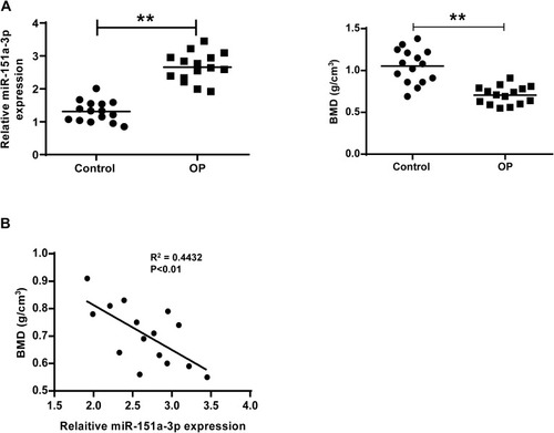
Figure 2 Overexpression of microRNA-151a-3p promoted RANKL-induced differentiation of THP-1 and RAW264.7 cells into osteoclasts. (A and B) RAW264.7 and THP-1 cells were treated with RANKL and M-CSF. (A) The representative images of TRAP-stained cells. Magnification, x200 (Day 0) and x400 (Day 7). (B) The expression levels of microRNA-151a-3p in RANKL/M-CSF-induced cells. (C and D) RANKL/M-CSF-induced RAW264.7 and THP-1 cells were treated with microRNA-151a-3p mimic. (C) Expression levels microRNA-151a-3p in RAW264.7 and THP-1 cells. (D) The expression levels of TRAP, NFATc1 and c-Fos. ** P < 0.01.
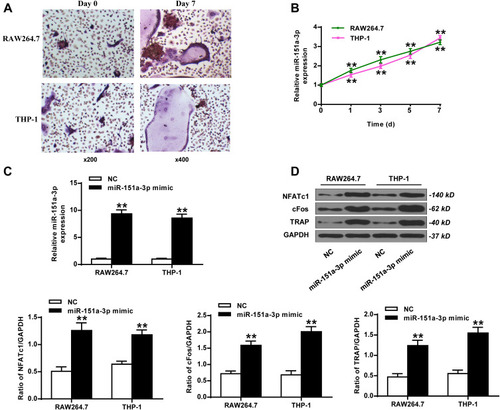
Figure 3 Silencing of microRNA-151a-3p restrained RANKL-induced differentiation of THP-1 cells and RAW264.7 cells into osteoclasts. RAW264.7 and THP-1 cells were treated with microRNA-151a-3p inhibitor. (A) The expression levels of microRNA-151a-3p in THP-1 cells and RAW264.7 cells. (B) The expression levels of TRAP, NFATc1 and c-Fos. **P < 0.01.
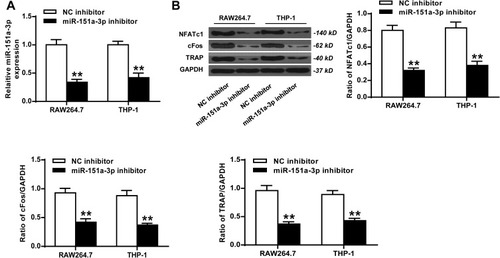
Figure 4 Silencing of microRNA-151a-3p altered the expression levels of osteoclastogenesis-related factors in sera of OVX rats. OVX rats were treated with antago microRNA-151a-3p or antago NC. (A) Expression levels of microRNA-151a-3p was detected in different groups. The expression levels of M-CSF (B), RANKL (B), TNF-α (C), IL-1α (C), CTX-I (D), and BGP (D) in different groups. N = 6 per group, *P < 0.05, **P < 0.01.
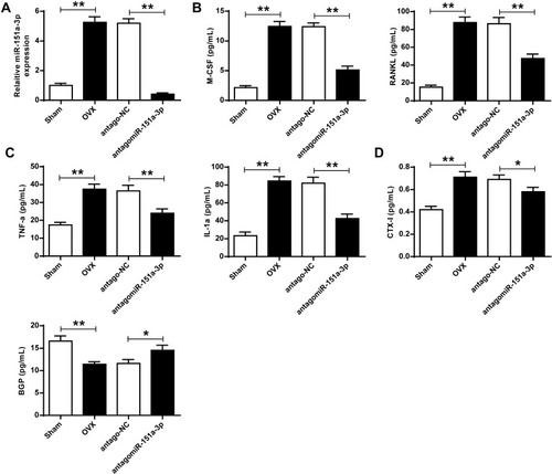
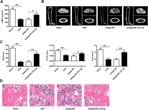
Figure 5 Knockdown of microRNA-151a-3p increased BMD and altered bone histomorphology in OVX rats. OVX rats were treated with antago microRNA-151a-3p or antago NC. (A) BMD was measured in different groups. (B and C) Bone histomorphometric parameters (BV/TV, Tb.Th, and Tb. N) were assessed. (D) Representative HE staining used to evaluate the pathological changes of osteogenic tissues in rats. 400×, Bar = 50 μm. N = 6 per group, *P < 0.05, **P < 0.01.