Figures & data
Table 1 General Information and Bone Density
Table 2 The Prevalence of Osteoporosis or Osteopenia in Different Age Groups
Table 3 BMI- and Gender-Related Distribution of Osteoporosis Diagnosed on DXA
Table 4 Diagnosis According to Lumbar DXA and Hip DXA
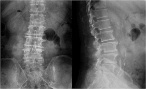
Figure 1 The anteroposterior and lateral X-ray images of a 68-year-old woman diagnosed with lumbar spinal stenosis. The T-scores of L1-L4 in the 68-year-old woman were −0.8, 0.4, 1.9 and 3.4, respectively. She had 3 abnormal segments with unreliable T-scores whose absolute value of T-score differences were all more than 1.0. From the anteroposterior and lateral X-ray images of the patient, we could observe obvious degenerative changes such as vertebral osteophytes and abdominal aortic calcifications, especially at lower segments.