Figures & data
Figure 1 Skin lesions. (A and B) Before treatment a large area of immersed erythema and edema with nodules, ulcerations, and crusts scattered on the right upper limb was present. A nummular deep ulcer with a severe tenderness presented on the back side of the second digit. (C and D) After 2 months of therapy the skin nodules decreased in size and the ulcer cavity was filled with fresh tissues. (E and F) After 6 months of therapy the lesions subsided, leaving some hyperplastic scars.
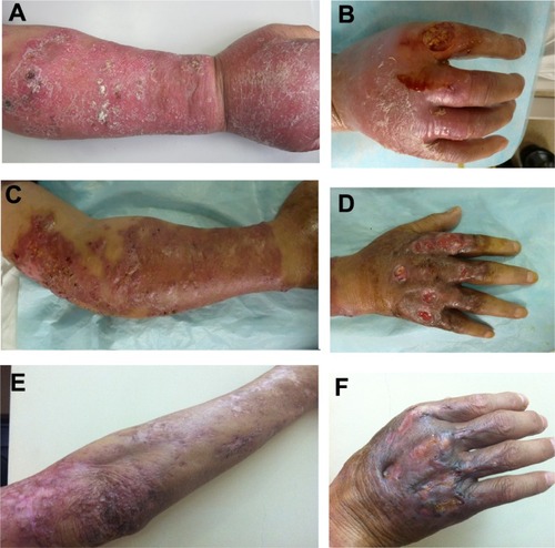
Figure 2 Pathological changes.
Note: A neutrophilic and histiocytic granuloma (hematoxylin and eosin; original magnification ×400).
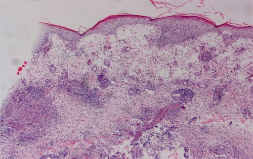
Figure 3 Pathogenic organism. (A) Mycobacterial cultures from the isolated lesional skin. (B) Smear from the cultured organisms with acid-fast stain.
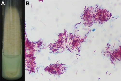
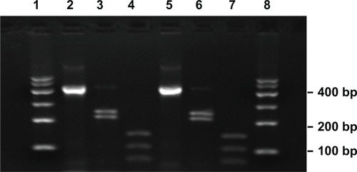
Figure 4 Restriction analyses of the isolated strain amplification products.