
Figures & data
Table 1 Characteristics of the Included Patients
Figure 1 Variation trends of radiographic and clinical indices in both the re-kyphosis and non-re-kyphosis group. Line graphs showing the changes in local kyphosis angle (LKA) (a), vertebral wedge angle (b), anterior intervertebral disc height (c), middle intervertebral disc height (d), posterior intervertebral disc height (e), visual analogue scale (VAS) scores (f) and Oswestry Disability Index (ODI) scores (g) of the two groups at three different time points.
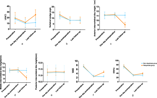
Figure 2 Typical case in the re-kyphosis group. A 67-year-old male patient complained of back pain for 10 days. T1-weighted (a), T2 -weighted (b) and fat-suppressed (c) MRI images showed bone marrow edema signal and disc–endplate complex injury of L1 vertebra(red arrow). Preoperative lateral X-ray image (d) showed multiple degenerative changes of lumbar spine. Lateral X-ray image 1 day after surgery (e) showed the vertebral height was restored and local kyphotic angle was corrected. Lateral X-ray image at the last follow-up (f) showed the occurrence of re-kyphosis.
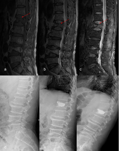
Table 2 Multivariate Logistic Regression Analysis