
Figures & data
Figure 1 Flowchart of patient eligibility screening.
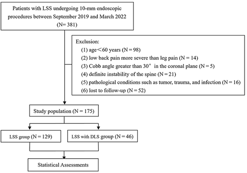
Figure 2 (A) Intraoperative anterior-posterior image. (B) Intraoperative lateral image. (C) Endoscope burr in use. (D) Removed compression-causing tissue.
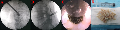
Table 1 Demographic Characteristics of the Two Groups
Table 2 Perioperative Indicators and Complications of the Two Groups
Figure 3 Clinical outcomes of patients in both groups at different follow-up time points. (A) VAS-back pain score (☆: P < 0.05). (B) VAS-leg pain score. (C) Oswestry Disability Index. (D) the Modified Macnab criteria.
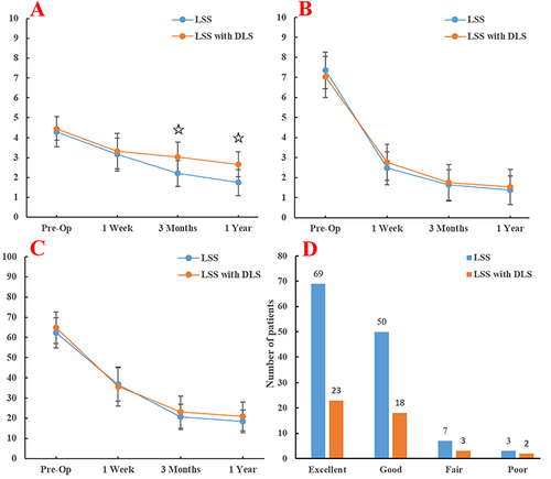
Table 3 Comparison of Imaging Parameters Between the Two Groups of Patients
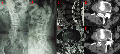
Figure 4 Imaging images of an 83-year-old DLS patient with L4/5 as the responsible segment. (A and B) X-rays showing degenerative deformity of the patient’s lumbar spine. (C and E) Preoperative lumbar MRI (Magnetic Resonance Imaging) showed severe spinal stenosis. (D and F) Axial CT (Computed Tomography) images of the lumbar spine show adequate decompression.
Data Sharing Statement
The data used to support the findings of this study are available from the corresponding author upon request.