Figures & data
Figure 1 Radiograph of the left foot, demonstrating a large lytic lesion and destruction of the calcaneus, the cuboid, and the fifth metatarsal.
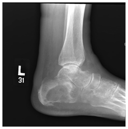
Figure 2 Left calcaneal biopsy specimen showing urothelial carcinoma with squamous differentiation.
Notes: Hematoxylin and eosin; magnification 100×.
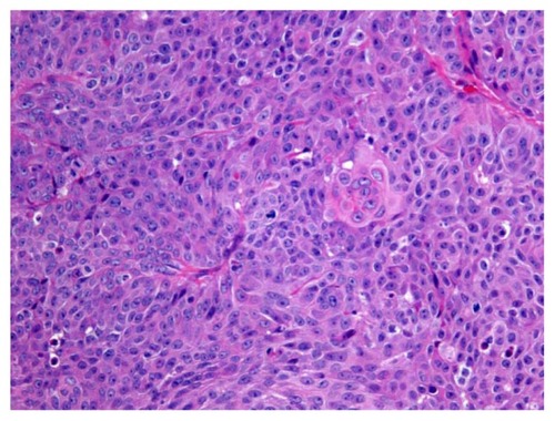
Figure 3 Abdominal and pelvic CT scan revealing a left hydronephrosis with renal cortical atrophy and an obstructing mass at the iliac crossing of the left ureter.
Abbreviation: CT, computed tomography.
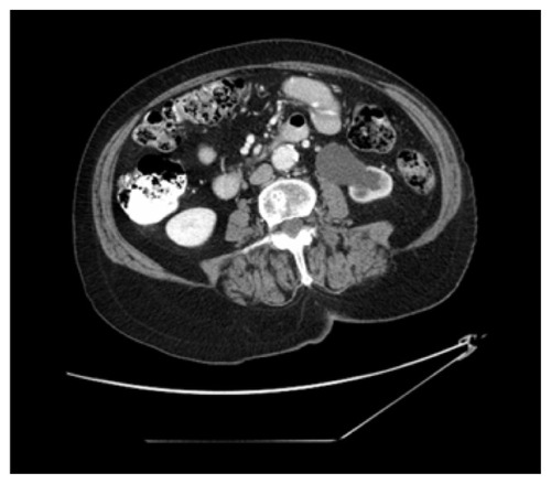
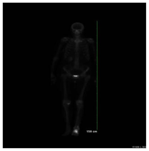
Figure 4 Technetium-99m MDP whole body bone scan displaying intense increased radioactive uptake in the left calcaneus with mild increased radioactive uptake in the proximal and distal left fibula with some extension into the shaft.
Abbreviation: MDP, methylene-diphosphonate.