Figures & data
Figure 1 Ulceration on the dorsal aspect of the left foot: inflammatory rim around the ulceration, desquamation, and diffuse erythema.
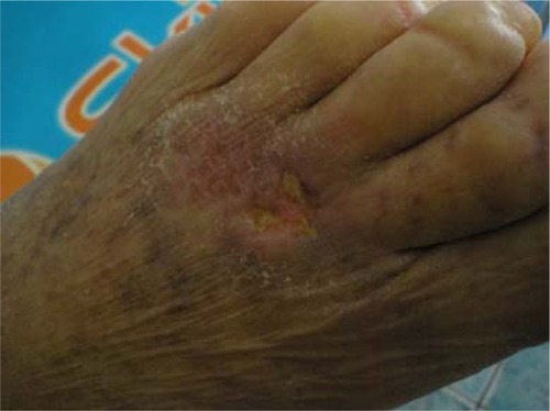
Figure 2 Proliferation of basaloid cell with palisading disposition (hematoxylin and eosin, 40×).
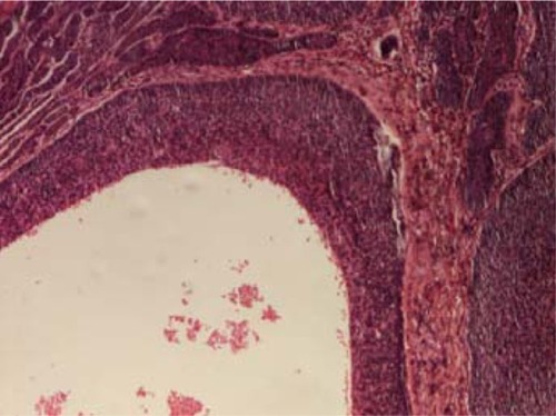
Figure 3 Clinical aspect after therapy: xerosis, small linear erosion, desquamation.
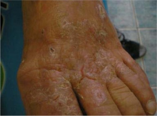
Figure 4 The tumoral lesion at admission.
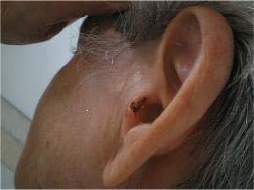
Figure 5 Islands of basaloid cells with palisading disposition in the upper dermis, with contraction around them (hematoxylin and eosin, 40×).
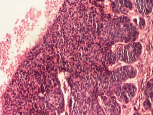
Figure 6 Clinical aspect after cryotherapy: no ulceration, no tumoral lesion, just a slight area of erythema.
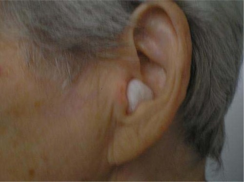
Figure 7 A small bleeding tumor covered by crust on the nose, multiple actinic keratosis on the face.
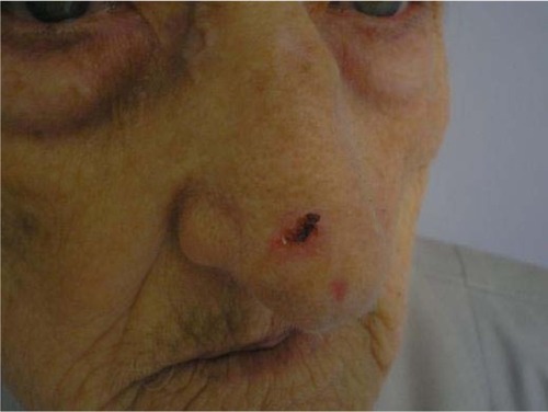
Figure 8 Basal cell carcinoma: typical nuclear palisading at the peripheral layer of the tumor (hematoxylin and eosin, 100×).
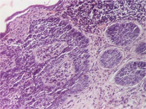
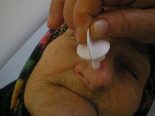
Figure 9 Cryotherapy procedure.