Figures & data
Figure 1 (A) Ropivacaine is injected into the epidural space using a spinal needle. (B) Dural sac and nerve root after decompression. LF, ligamentum flavum; NR, nerve root.
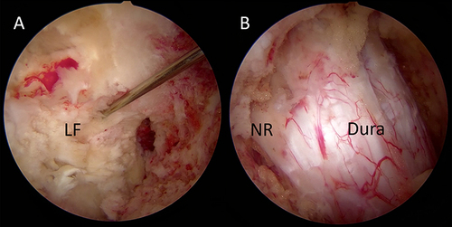
Table 1 Patient Demographic and Clinical Characteristics
Table 2 Surgical Variables
Table 3 Patient-Reported Outcomes Measures
Figure 2 Clinical outcomes based on the modified Macnab criteria at last follow-up.
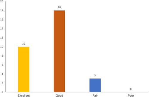
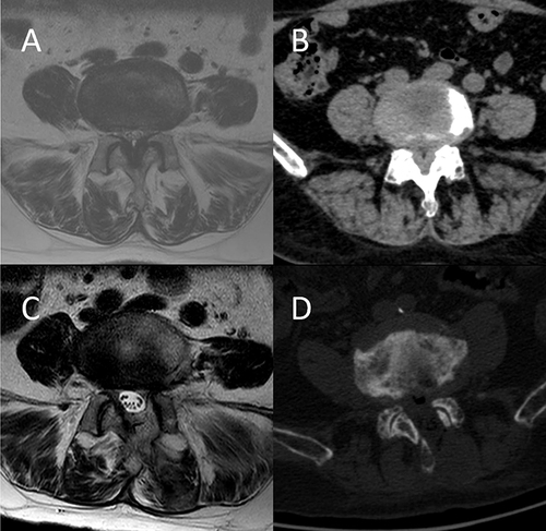
Figure 3 Typical case: A 75-year-old female patient complained of neurogenic claudication and was diagnosed with L4-5 degenerative lumbar spinal stenosis. (A and B) Preoperative axial MR and CT images at L4-5. (C and D) Postoperative axial MR and CT images.
Abbreviations: MR, magnetic resonance; CT, computed tomography.