Figures & data
Table 1 Clinical Features of SGD Group and Non-SGD Group
Table 2 Clinical Features of Ulcer Group and Non-Ulcer Group (Brownish Scaly Patch and Erythema)
Figure 1 Lesions of senile gluteal dermatosis (A) Thickened brownish scaly patches on gluteal cleft apex and both sides of the buttocks form “three corners of a triangle” with distinct horizontal ridges; (B): brownish scaly patches, erythema and shallow ulcers were seen in one patient; (C): A crusted ulcer can be seen on the skin corresponding to the greater trochanter of the femur.
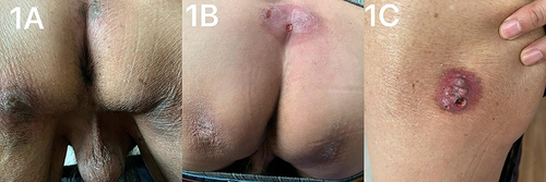
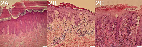
Figure 2 Pathological findings of SGD. (A) The brownish scaly plaque patients showed significant hyperkeratosis, some with epidermal psoriatic hyperplasia, and perivascular lymphocyte infiltration. (B) The pathological manifestations of erythema patients include mild hyperkeratosis, focal hyperkeratosis, focal mild spongy edema of the epidermis, and small to moderate amount of lymphocyte infiltration around superficial vessels of the dermis; (C) Pseudoepitheliomatous hyperplasia and massive perivascular lymphocyte infiltration were seen in patients with ulcers.