
Figures & data
Figure 1 Physiologic and Pathologic Images of the Retina. (a) Structure diagram of retinal layers and immunosuppressive mechanism in the retina; (b) Pathological changes in dry AMD: drusen formation, RPE dysfunction; (c) Pathological changes in wet AMD: CNV formation, photoreceptor/RPE/BM/CC atrophy and dysfunction.
Note: Created with BioRender.com.
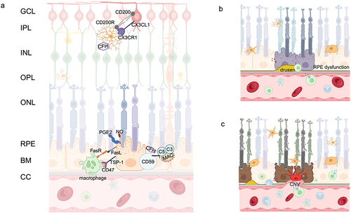
Figure 2 Vicious Cycle. Mechanisms of interaction between cellular senescence and dysregulation of immune homeostasis in retina.
Note: Created with BioRender.com.
