Figures & data
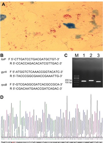
Figure 1 (A) Microscopic observation of numerous acid fast bacilli (red rods) in the slit skin smear fluid from a solitary lesion in the face at the initial presentation of the patient. Original magnification: ×1,000. (B) primers used in this study. The letters F and R refer to forward and reverse primer, respectively. (C) Amplification products of folP, gyrA, and rpoB genes (lanes 1–3, respectively). (D) A mutation of codon 441 of the rpoB gene was found (GAt→AAC) (indicated in red).