Figures & data
Figure 1 The AxiaLIF rod.
Note: 50 mm length shown.
Abbreviation: AxiaLIF, axial lumbar interbody fusion.
Abbreviation: AxiaLIF, axial lumbar interbody fusion.
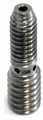
Figure 2 Presacral access with the AxiaLIF system.
Notes: Dilator and guide pin advanced into the L5–S1 interspace (A). Radial cutters debulking disc material and preparing the endplates for fusion (B). Insertion of bone graft material in the L5–S1 interspace (C). AxiaLIF rod implantation to within 5–10 mm of superior endplate of L5 (D). Images provided courtesy of Baxano surgical, Inc.Citation32
Abbreviation: AxiaLIF, axial lumbar interbody fusion.
Abbreviation: AxiaLIF, axial lumbar interbody fusion.
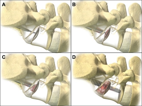
Table 1 Baseline patient characteristics
Table 2 Univariate baseline predictors of solid fusion at final postoperative follow-up
Figure 3 Preoperative T1 (A) and T2 (B) images of a 34-year-old woman with a 6-year history of back pain showing discopathy at the L5/S1 level with Modic 1 endplate changes. Postoperative sagittal (C) and coronal (D) CT at 1 year demonstrates solid fusion.
Abbreviation: CT, computed tomography.
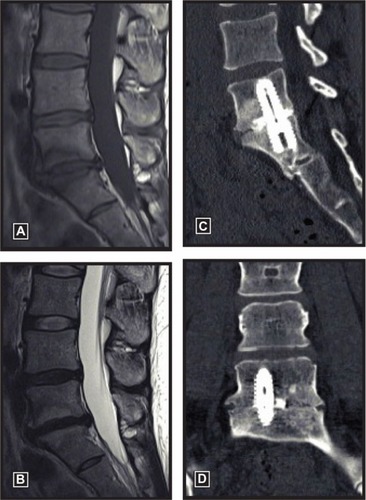
Table 3 Comparison of studies with axial lumbar interbody fusion for degenerative disc disease