Figures & data
Figure 1 Fluoroscopic image showing radiofrequency cannula toward the articular branches of the femoral nerve (A) and the obturator nerve (B).
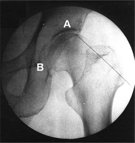
Table 1 Demographics of both study groups
Table 2 Comparison of outcomes between both study groups
Figure 2 CONSORT diagram for patient recruitment, allocation, and follow-up.
Abbreviations: CONSORT, Consolidated Standards of Reporting Trials; PRF, pulsed radiofrequency.
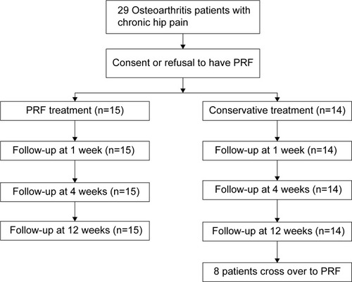
Figure 3 Longitudinal changes in mean (± SD) VAS scores.
Notes: Patients treated by PRF are compared with patients treated conservatively. Asterisk indicates P<0.05 for between-group comparisons.
Abbreviations: PRF, pulsed radiofrequency; SD, standard deviation; VAS, visual analog scale.
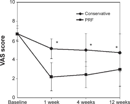
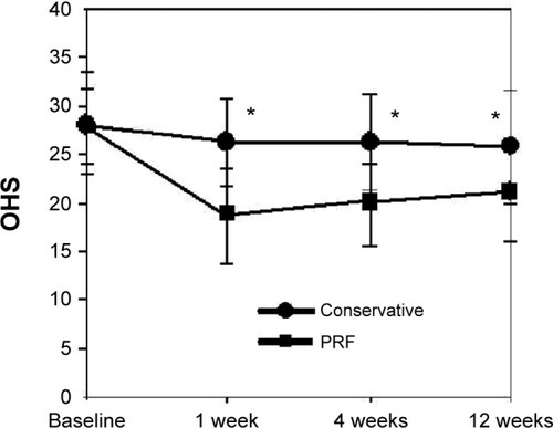
Figure 4 Longitudinal changes in mean (± SD) Oxford hip scores.
Notes: Patients treated by PRF are compared with patients treated conservatively. Asterisk indicates P<0.05 for between-group comparisons.
Abbreviations: OHS, Oxford hip scores; PRF, pulsed radiofrequency; SD, standard deviation.
Abbreviations: OHS, Oxford hip scores; PRF, pulsed radiofrequency; SD, standard deviation.