Figures & data
Figure 1 Effects of three antiarthritics on body weight of rats.
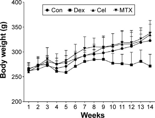
Table 1 Effects of three antiarthritics treatments on serum biochemical indicators of rats
Table 2 Effects of three antiarthritics on histomorphometric static parameters of the proximal tibial metaphysis
Table 3 Effects of three antiarthritics on bone formation parameters and osteoclast surface of proximal tibial metaphysis
Figure 2 Representative micrographs of the proximal tibial metaphysis.
Abbreviations: Con, saline control; Dex, dexamethasone; Cel, celecoxib; MTX, methotrexate.
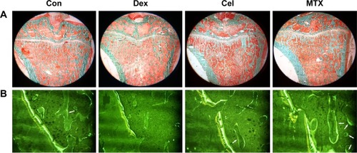
Figure 3 Effects of three antiarthritics on micro-CT parameters of the proximal tibial metaphysis.
Abbreviations: Con, saline control; Dex, dexamethasone; Cel, celecoxib; MTX, methotrexate; micro-CT, micro-computed tomograph; TV, total tissue volume.
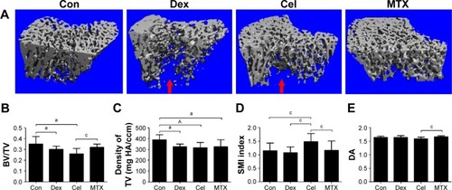
Figure 4 Effects of three antiarthritics on BMD of the femur and the fifth lumbar vertebrae in rats.
Abbreviations: Con, saline control; Dex, dexamethasone; Cel, celecoxib; MTX, methotrexate; BMD, bone mineral density.
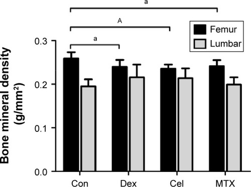
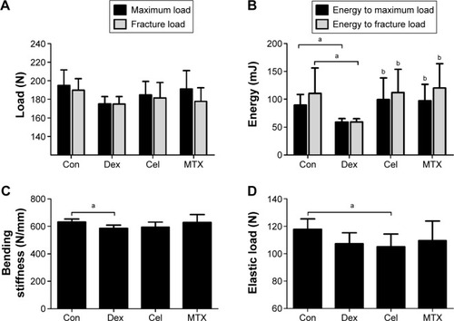
Figure 5 Effects of three antiarthritics on bone biomechanical parameters of femur in rats.
Abbreviations: Con, saline control; Dex, dexamethasone; Cel, celecoxib; MTX, methotrexate.