Figures & data
Figure 1 Chromatographic fingerprint analysis and SH-SY5Y cell viabilities of the herbal formula B307.
Abbreviations: AU, arbitrary perfusion units; HPLC, high-performance liquid chromatography; RA, retinoic acid.
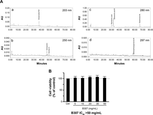
Figure 2 Life span, body weight, and motor ability of R6/2 HD mice and their WT under oral B307 and sham treatments.
Abbreviations: WT, wild-type littermate controls; HD, Huntington’s disease; H&E, hematoxylin and eosin; mHtt, mutant huntingtin; IHC, immunohistochemical; SEM, standard error of the mean; ANOVA, analysis of variance.
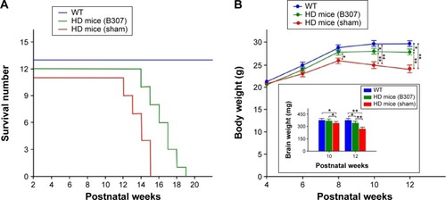
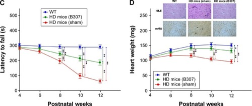
Figure 3 Cardiac performance in R6/2 HD mice under oral B307 and sham treatments, and of their WT.
Abbreviations: WT, wild-type littermate controls; HD, Huntington’s disease; MV, mitral valve; AV, aortic valve; SEM, standard error of the mean; ANOVA, analysis of variance.
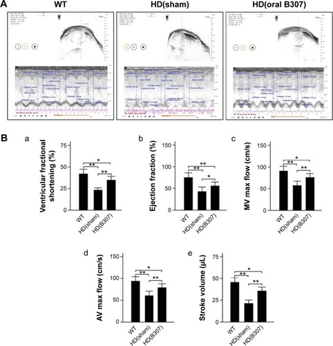
Figure 4 Expressions of 3-NT and 4-HNE, two markers of oxidative stress, and SOD2, a marker of antioxidative stress, in the heart tissue of R6/2 HD mice under oral B307 treatment, and of their WT.
Abbreviations: WT, wild-type littermate controls; HD, Huntington’s disease; 3-NT, neurotrophin-3; SOD2, superoxide dismutase 2; 4-HNE, 4-hydroxynonenal; IHC, immunohistochemical; SEM, standard error of the mean; ANOVA, analysis of variance.
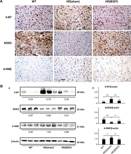
Figure 5 Expression of TNF-α, a marker of inflammation, in the heart tissue of R6/2 HD mice under oral B307 treatment, and of their WT.
Abbreviations: WT, wild-type littermate controls; HD, Huntington’s disease; TNF-α, tumor necrosis factor alpha; IHC, immunohistochemical; SEM, standard error of the mean; ANOVA, analysis of variance.
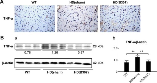
Figure 6 Expressions of Bcl-2, a marker of anti-apoptosis, and Bax and Cyto-C, two markers of apoptosis, in the heart tissue of R6/2 HD mice under oral B307 treatment.
Notes: (A) IHC staining illustrates that expressions of Bcl-2 (shown in brown) were weaker than their WT but were enhanced under oral B307 treatment. Further expressions of Bax and Cyto-C (shown in brown) were remarkable in comparison to their WT but were reduced under oral B307 treatment. Scale bars: 25 μm. (B) Western blotting analysis shows the following: (a) Cardiac expression levels of Bcl-2, Bax, and Cyto-C in R6/2 HD mice under sham and oral B307 treatments, and of their WT. (b) Quantified cardiac Bcl-2 levels in R6/2 HD mice were significantly weaker than their WT but were significantly enhanced under oral B307 treatment. Further quantified cardiac Bax and Cyto-C levels in R6/2 HD mice were significantly enhanced in comparison to their WT but were significantly weaker under oral B307 treatment. There were six mice per each group. Values are mean ± SEM (**P<0.01, *P<0.05, two-way ANOVA followed by a Student–Newman–Keuls multiple comparisons posttest).
Abbreviations: WT, wild-type littermate controls; HD, Huntington’s disease; Bcl-2, B-cell lymphoma 2; Bax, Bcl-2-associated X protein; Cyto-C, cytochrome C; IHC, immunohistochemical; SEM, standard error of the mean; ANOVA, analysis of variance.
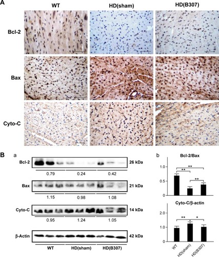
Figure 7 Expressions of calpain and caspase 12, two markers of ER stress-related apoptosis, in the heart tissue of R6/2 HD mice under oral B307 treatment, and of their WT.
Notes: (A) IHC staining illustrates that expressions of calpain and caspase 12 (shown in brown) were remarkable in comparison to their WT but were reduced under oral B307 treatment. Scale bars: 25 μm. (B) Western blotting analysis shows the following: (a) Cardiac expression levels of calpain and caspase 12 in R6/2 HD mice under sham and oral B307 treatments, and of their WT. (b) Quantified cardiac calpain and caspase 12 levels in R6/2 HD mice were significantly enhanced in comparison to their WT but were significantly weaker under oral B307 treatment. There were six mice per each group. Values are mean ± SEM (**P<0.01, *P<0.05, two-way ANOVA followed by a Student–Newman–Keuls multiple comparisons posttest).
Abbreviations: WT, wild-type littermate controls; HD, Huntington’s disease; ER, endoplasmic reticulum; IHC, immunohistochemical; SEM, standard error of the mean; ANOVA, analysis of variance.
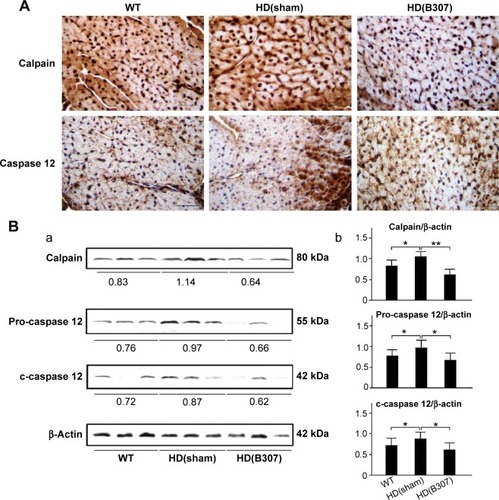
Figure 8 Expressions of caspase 9 and caspase 3, two markers of apoptosis, in the heart tissue of R6/2 HD mice under oral B307 treatment and of their WT.
Abbreviations: WT, wild-type littermate controls; HD, Huntington’s disease; IHC, immunohistochemical; SEM, standard error of the mean; ANOVA, analysis of variance.
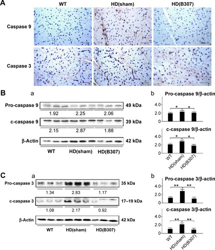
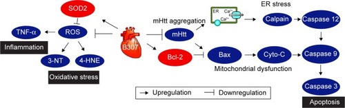
Figure 9 The schematic diagram illustrates the possible cardioprotective pathways under oral B307 treatment.
Abbreviations: mHtt, mutant huntingtin; SOD2, superoxide dismutase 2; ROS, reactive oxygen species; 3-NT, neurotrophin-3; 4-HNE, 4-hydroxynonenal; TNF-α, tumor necrosis factor alpha; ER, endoplasmic reticulum; Bcl-2, B-cell lymphoma 2; Bax, Bcl-2-associated X protein; Cyto-C, cytochrome C; ER, endoplasmic reticulum; WT, wild-type littermate controls; HD, Huntington’s disease.