Figures & data
Table 1 Electrophysiological features of the patient were comparable with typical length-dependent, predominantly axonal sensory-motor polyneuropathy
Figure 1 Electrocardiogram revealed sinus rhythm, low voltages in limb leads, QS waves indicative of pseudoinfarction in precordial and inferior leads, first-degree atrioventricular block, and prolonged QTc.
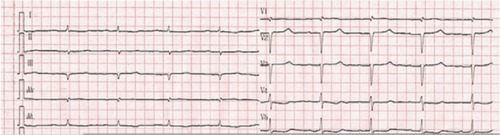
Figure 2 A four-chamber apical view echocardiogram showing biatrial dilatation, valve thickening, thick ventricular walls (left ventricular wall is 15 mm and interventricular septum is 19 mm), and interventricular septum with speckled appearance, which suggests amyloid infiltrate.

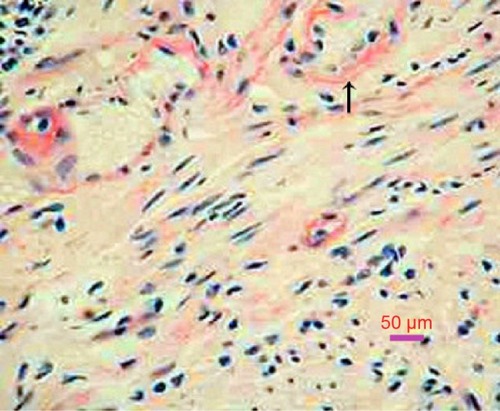
Figure 3 Rectum biopsy: amyloid deposits are confirmed by a positive Congo red stain (arrow), which gives the characteristic salmon pink color (200×).