Figures & data
Figure 1 Electrocardiogram on admission and after stent implantation.
Notes: (A) Showing ST segment slope-down depression and T-wave inversion in leads II, III, aVF, and V4–V6; (B) ECG after stent implantation. T-wave amplitudes in leads II, III, aVF were decreased, compared with (A).
Abbreviation: ECG, electrocardiogram.
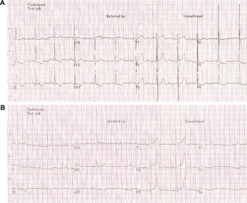
Figure 2 Colon under colonoscopy.
Notes: (A, B) Colonoscopy showing tumor with dirty white-yellow furs in the transverse colon. (C, D) Repeat colonoscopy showing normal colon 10 months after the operation.
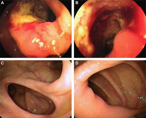
Figure 3 Coronary angiogram and PCI.
Notes: (A) Diffuse stricture (70%–85%) of proximal and middle circumflex and focal proximal left anterior descending stenosis 60% on right anterior oblique projection; (B) stenosis on left anterior oblique projection; (C) middle right coronary occlusion, 95% stenosis of the opening of the first sharp edge of branch on right anterior oblique projection; (D) right coronary artery on right anterior oblique after stent implantation; (E) stent thrombosis of right coronary artery on right anterior oblique; (F, G) inflated balloon in proximal and distal parts of the stent during PTCA; (H) right coronary artery after PTCA on right anterior oblique projection.
Abbreviations: PCI, percutaneous coronary intervention; PTCA, percutaneous transluminal coronary angioplasty.
Abbreviations: PCI, percutaneous coronary intervention; PTCA, percutaneous transluminal coronary angioplasty.
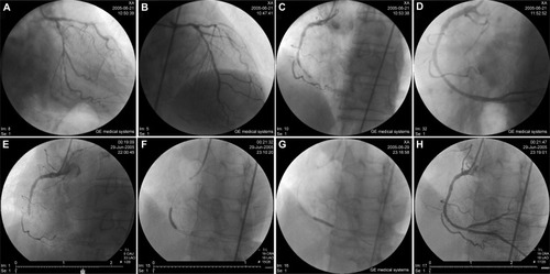
Figure 4 ECG during stent thrombosis.
Notes: (A) ECG before recurrent chest pain. (B) Inverted T-waves in leads II, III, and aVF. (C) Increased T-wave amplitudes in leads III, aVF, and V1–V6, compared with (B); (D) ECG after PTCA, decreased T-wave amplitudes in leads II, III, and aVF, compared with (C).
Abbreviations: ECG, electrocardiogram; PTCA, percutaneous transluminal coronary angioplasty.
Abbreviations: ECG, electrocardiogram; PTCA, percutaneous transluminal coronary angioplasty.
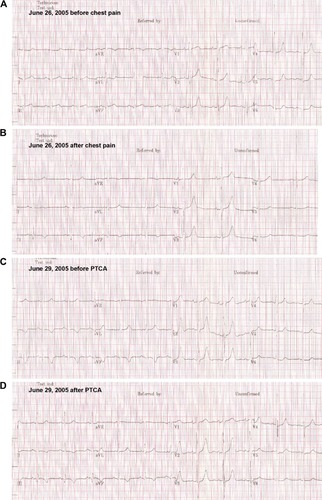
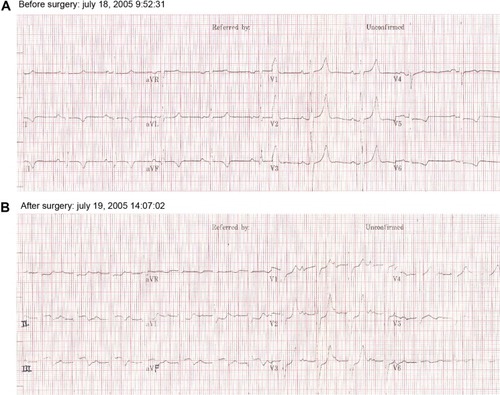
Figure 5 ECG changes of acute inferior wall myocardial infarction post-operation.
Notes: (A) ECG 1 day before surgery; (B) obvious elevation of ST segment in leads II and III, aVF compared with (A).
Abbreviation: ECG, electrocardiogram.
Abbreviation: ECG, electrocardiogram.