Figures & data
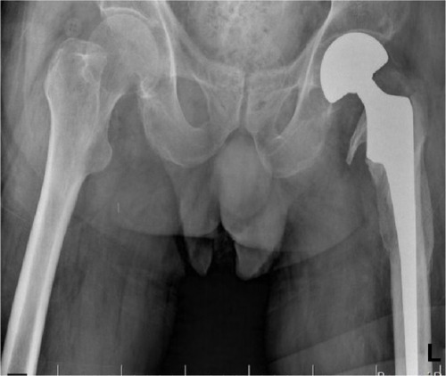
Figure 1 Radiographs showing the periprosthetic destructive area at femoral medial cortex below lesser trochanter.
Abbreviation: L, left.
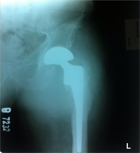
Figure 2 Angiogram showing the pseudoaneurysm was detected in profunda branch of femoris artery and periprosthetic destructive area.
Abbreviation: L, left.
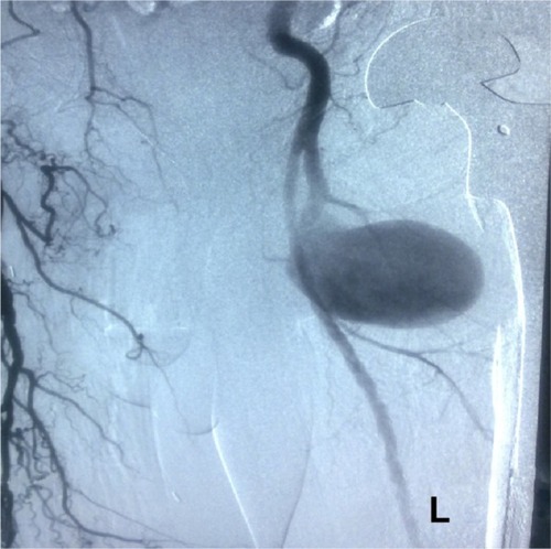
Figure 3 Radiographs showing the remodeling initiated in osteolysis site at month 2 after surgery.
Abbreviation: L, left.
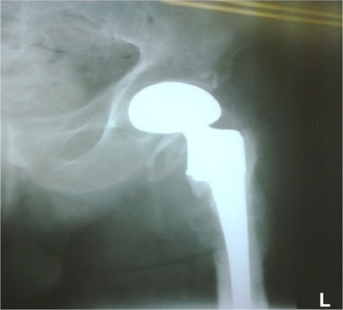
Figure 4 On the control visit at year 2, it was seen that the osteolytic area had completely recovered.
Abbreviation: L, left.