Figures & data
Figure 1 Intraoperative small hemorrhages during manual dilation of small pupil.
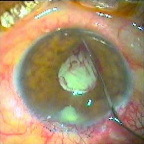
Figure 2 Small iris sphincter ruptures.
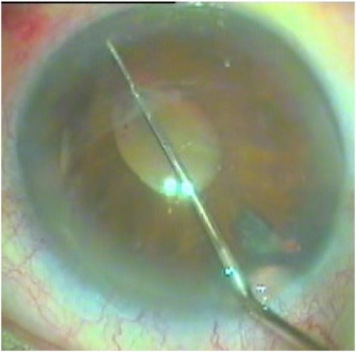
Table 1 Patient demographics
Table 2 Group I mean pupil size
Table 3 Corneal edema and iritis/anterior chamber flare at the first postoperative day and BCVA on the 30th postoperative day
Figure 3 Comparison of postoperative corneal edema between the two groups, P=0.92.
One month after surgery, the pupils were round and reactive to light; the anterior chambers were quiet; and the corneas were clear in all studied eyes. The BCVA on Snellen chart was 20/40 (Monoyer’s scale) or better and there were no statistically significant differences between the two groups (–).
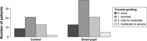
Figure 4 Comparison of postoperative iritis between the two groups, P=0.25.
Abbreviation: ac, anterior chamber.
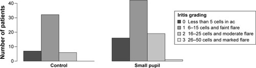
Figure 5 Comparison of postoperative VA between the two groups, P=0.32.
Abbreviations: BCVA, best-corrected visual acuity; VA, visual acuity.
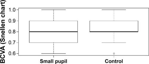
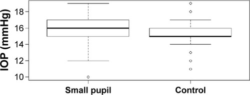
Figure 6 Comparison of postoperative IOP between the two groups, P=0.11.