
Figures & data
Figure 1 Study design.
Notes: The source population consisted of all female residents aged 35–69 of Denmark’s Jutland Peninsula between 1985 and 2001, who were diagnosed with non-metastatic breast cancer. Two-thirds of the women (n = 7617) were excluded because of an unknown treatment protocol or because they did not meet the inclusion criteria. Ki-67 results were missing if tissue was unavailable or if the tumor core was unsatisfactory after processing, staining, and imaging.
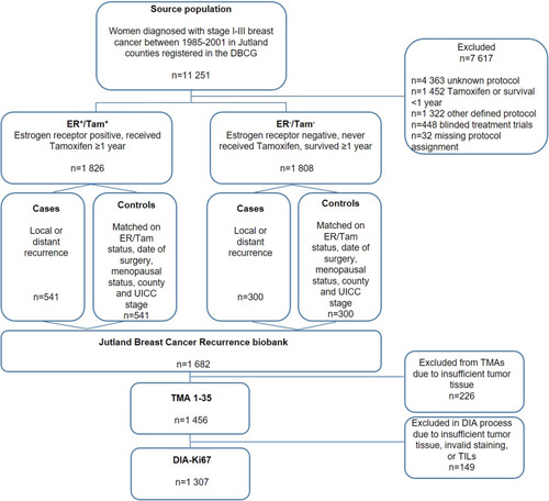
Figure 2 Tumor region of interest (ROI).
Notes: ROI (outlined in green) was defined semi-automatically in Visiopharm®, based on both size and morphology of the cells. Stroma and TILs were disregarded by the customized APP.
Abbreviations: APP, analysis protocol package; TILs, tumor-infiltrating lymphocytes.
Abbreviations: APP, analysis protocol package; TILs, tumor-infiltrating lymphocytes.
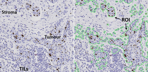
Figure 3 Representation of DIA scoring of Ki-67.
Notes: (A) before, and (B) after, the customized algorithm was run in the Visiopharm® program. Ki-67-positive tumor cells were identified and scored in relation to the negative tumor cells: Ki-67 positively stained nuclei were identified based on their brown DAB staining, whereas negative cells were identified based on their blue H&E stain. Ki-67 score= [(area of Ki-67-positive tumor cells)/(area positive + negative tumor cells) x 100]. DIA score in this particular core was calculated by the customized algorithm to be 61%.
Abbreviation: DIA, digital image analysis.
Abbreviation: DIA, digital image analysis.
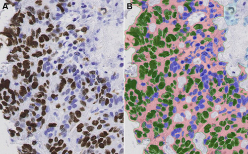
Table 1 Patient and Clinical Characteristics for Cases and Controls of the Jutland Breast Cancer Recurrence Biobank
Table 2 Associations Between Ki-67 Expression and Breast Cancer Recurrence Within ER/Tam Groups