Figures & data
Figure 1 RRBP1 expression.
Notes: The expression of RRBP was detected in esophageal carcinoma and matched adjacent normal tissues by qRT-PCR (A) and Western blot (B). T, esophageal carcinoma tissue; N, matched adjacent normal esophageal tissue.
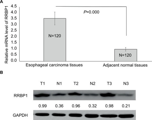
Table 1 RRBP1 expression in esophageal carcinoma and normal esophageal tissues by immunohistochemical staining
Table 2 RRBP1 expression correlation with clinicopathological characteristics in esophageal carcinoma
Figure 2 RRBP1 expression was detected in esophageal carcinoma and matched adjacent normal tissues by immunohistochemical staining.
Notes: (A) Adjacent normal tissues; (B) weak staining of RRBP1 in esophageal carcinoma; (C) moderate staining of RRBP1 in esophageal carcinoma; (D) strong staining of RRBP1 in esophageal carcinoma.
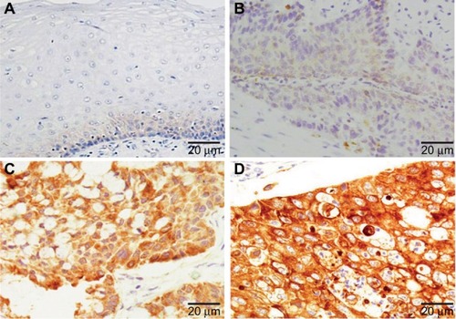
Table 3 Patient survival: Kaplan–Meier survival analysis
Table 4 Patients’ survival evaluation by multivariate Cox regression analysis
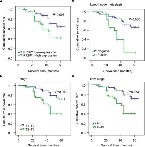
Figure 3 Kaplan–Meier survival analysis.
Notes: Results indicated that RRBP1 expression (A), lymph node metastasis (B), T stage (C), and TNM (D) stage were associated with patients’ prognosis.