
Figures & data
Table 1 Summary of the histological features of orbital SFT in the present series
Figure 1 Histopathological and immunohistochemical staining of tumors in our cases.
Notes: (A) The tumor is composed of haphazardly arranged spindle cells with bland nuclei and inconspicuous nucleoli. (Case 1; HE staining; original magnification: ×200). (B) Variably collagenous and numerous endothelium-lined vascular channels are noted. (Case 2; arrow shows an occasional vascular channel with a staghorn appearance; HE staining; original magnification: ×200). (C) Tumor cells with oval nuclei, conspicuous nucleoli, and mitotic figure are noted. (recurrent sample of Case 4; HE staining; original magnification: ×200). (D) Tumor cells are positive for CD34. (recurrent sample of Case 4; original magnification: ×200). (E) The tumor cells are positive for Bcl-2 (Case 2; original magnification: ×200). (F) The tumor cells are positive for Ki-67 (>10%), which indicates malignant transformation (recurrent sample of Case 4; original magnification: ×200).
Abbreviations: HE, hematoxylin and eosin; CD34, cluster of differentiation 34; Bcl-2, B-cell lymphoma 2.
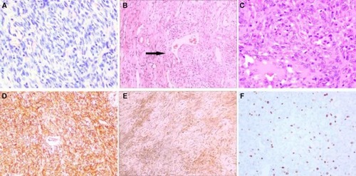
Figure 2 MR scan of Case 2 shows a well-circumscribed circular mass in the subcutaneous region of the right lacrimal sac area.
Notes: (A) T1 image, (B) T2 image, and (C) post-contrast T1 image.
Abbreviation: MR, magnetic resonance.
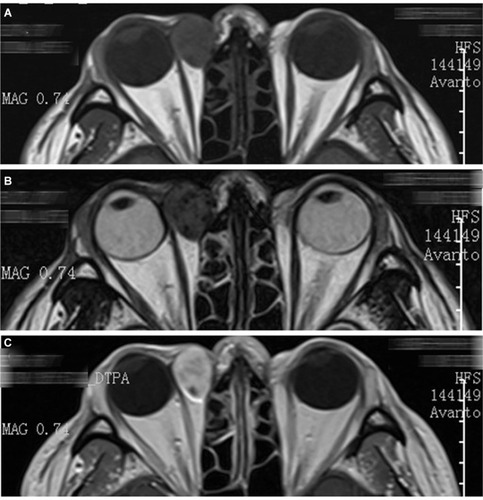
Figure 3 MR scan of Case 3 shows a well-circumscribed subcutaneous mass in the superomedial side of her left orbit with homogeneous isointense signal on T1 images and hypointense signal on T2 images.
Notes: (A) T1 image, (B) T2 image, and (C) post-contrast T1 image.
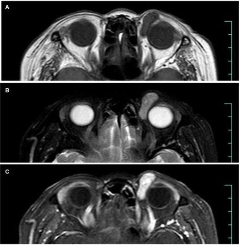
Figure 4 The MR scan of Case 4 (1st op). Arrows show the lesion had two types of imaging features on MRI. The medial part of the mass indicates a tissue with high collagen content whereas the lateral part indicates a tissue with hypervascular content.
Notes: (A, D) T1 image, (B, E) T2 image, and (C, F) post-contrast T1 image.
Abbreviation: op, operation.
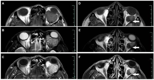
Figure 5 MR scan of Case 4 (2nd operation). Arrows show signal tubular structures, which might represent fast-flow vessels within the tumor.
Notes: (A, D) T1 image, (B, E) T2 image, and (C, F) post-contrast T1 image.
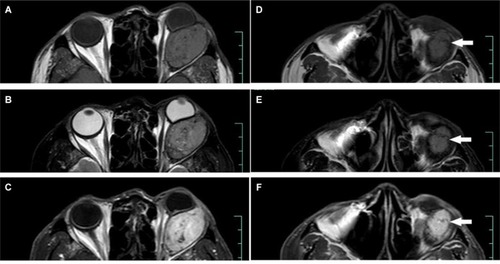
Table 2 Summary of clinical features of orbital SFT in the present series
Table 3 Summary of reported recurrent orbital SFT with malignant transformation